See how this article has been cited at scite.ai
scite shows how a scientific paper has been cited by providing the context of the citation, a classification describing whether it supports, mentions, or contrasts the cited claim, and a label indicating in which section the citation was made.
Combined lectin- and immuno-histochemistry (CLIH) for applications in cell biology and cancer diagnosis: Analysis of human urothelial carcinomas
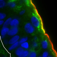
Lectin histochemistry (LHC) and immunohistochemistry (IHC), which demonstrate the composition and localisation of sugar residues and proteins in cell membranes, respectively, are generally used separately. Using these two methods, we previously demonstrated that malignant transformation of urothelial cells results in the alterations of protein glycosylation and reduced expression of urothelium-specific integral membrane proteins uroplakins (UPs). However, the correlation between these changes was not studied yet. To evaluate this correlation, we developed innovative method, which we named combined lectin- and immuno- histochemistry (CLIH). We used human biopsies of 6 normal urothelia and 9 papillary urothelial carcinomas, i.e. 3 papillary urothelial neoplasms of low malignant potential (PUNLMP), 3 non-invasive papillary urothelial carcinomas of low grade (pTa, l.g.), and 3 invasive papillary urothelial carcinomas of high grade (pT1, h.g.). We tested five different protocols (numbered 1-5) of CLIH on paraffin and cryo-semithin sections and compared them with LHC and IHC performed separately. Additionally, we carried out western and lectin blotting with antibodies against UPs and lectins Amaranthus caudatus agglutinin (ACA), Datura stramonium agglutinin (DSA), and jacalin, respectively. We showed that incubation with primary antibodies first, followed by the mixture of secondary antibodies and lectins is the most efficient CLIH method (protocol number 5). Additionally, 300 nm thick cryo-semithin sections enabled better resolution of co-localisation between sugar residues and proteins than 5 µm thick paraffin sections. In the normal urothelium, CLIH showed co-localisation of lectins ACA and jacalin with UPs in the apical plasma membrane (PM) of superficial umbrella cells. In papillary urothelial carcinomas, all three lectins (ACA, DSA and jacalin) labelled regions of apical PM, where they occasionally co-localised with UPs. Western and lectin blotting confirmed the differences between normal urothelium and papillary urothelial carcinomas. Our results show that CLIH, when used with various sets of lectins and antigens, is a useful, quick, and reliable method that could be applied for basic cell biology research as well as detailed subtyping of human urothelial carcinomas.


Supporting Agencies
Slovene Research Agency (research core funding No. P3-0108 and Infrastructure program MRIC UL IP-0510)How to Cite
PAGEPress has chosen to apply the Creative Commons Attribution NonCommercial 4.0 International License (CC BY-NC 4.0) to all manuscripts to be published.