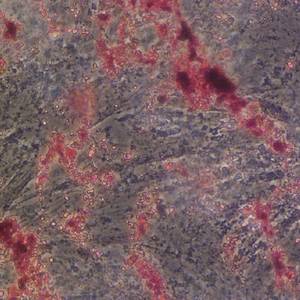
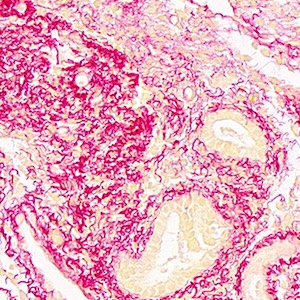
Immunohistochemical detection of early-stage carcinogenesis of oral leukoplakia by increased DNA-instability and various malignancy markers
Submitted: 29 December 2009
Accepted: 29 December 2009
Published: 30 December 2009
Accepted: 29 December 2009
Abstract Views: 575
PDF: 719
Publisher's note
All claims expressed in this article are solely those of the authors and do not necessarily represent those of their affiliated organizations, or those of the publisher, the editors and the reviewers. Any product that may be evaluated in this article or claim that may be made by its manufacturer is not guaranteed or endorsed by the publisher.
All claims expressed in this article are solely those of the authors and do not necessarily represent those of their affiliated organizations, or those of the publisher, the editors and the reviewers. Any product that may be evaluated in this article or claim that may be made by its manufacturer is not guaranteed or endorsed by the publisher.
Authors
PAGEPress Office, Pavia, Italy.
Downloads
Download data is not yet available.
Publication Facts
Metric
This article
Other articles
Peer reviewers
0
2.4
Reviewer profiles N/A
Author statements
Author statements
This article
Other articles
Data availability
N/A
16%
External funding
N/A
32%
Competing interests
N/A
11%
Metric
This journal
Other journals
Articles accepted
57%
33%
Days to publication
0
145
- Editor & editorial board
-
profiles
- Academic society
- N/A
- Publisher
- PAGEPress Publications, Pavia, Italy
To learn about these publication facts, click
PF is maintained by the Public Knowledge Project
How to Cite
Iwasa, M., Imamura, Y., Noriki, S., Nishi, Y., Kato, H., & Fukuda, M. (2009). Immunohistochemical detection of early-stage carcinogenesis of oral leukoplakia by increased DNA-instability and various malignancy markers. European Journal of Histochemistry, 45(4), 333–46. https://doi.org/10.4081/1642
PAGEPress has chosen to apply the Creative Commons Attribution NonCommercial 4.0 International License (CC BY-NC 4.0) to all manuscripts to be published.

 https://doi.org/10.4081/1642


