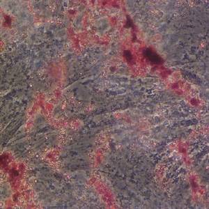
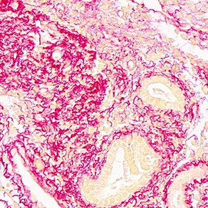
Simultaneous staining of cytoplasmic iron and collagen matrix in human liver biopsy specimens
Submitted: 30 December 2009
Accepted: 30 December 2009
Published: 30 December 2009
Accepted: 30 December 2009
Abstract Views: 469
PDF: 468
Publisher's note
All claims expressed in this article are solely those of the authors and do not necessarily represent those of their affiliated organizations, or those of the publisher, the editors and the reviewers. Any product that may be evaluated in this article or claim that may be made by its manufacturer is not guaranteed or endorsed by the publisher.
All claims expressed in this article are solely those of the authors and do not necessarily represent those of their affiliated organizations, or those of the publisher, the editors and the reviewers. Any product that may be evaluated in this article or claim that may be made by its manufacturer is not guaranteed or endorsed by the publisher.

 https://doi.org/10.4081/1658


