Overexpression of kynurenic acid and 3-hydroxyanthranilic acid after rat traumatic brain injury

Accepted: 2 November 2018
HTML: 16
All claims expressed in this article are solely those of the authors and do not necessarily represent those of their affiliated organizations, or those of the publisher, the editors and the reviewers. Any product that may be evaluated in this article or claim that may be made by its manufacturer is not guaranteed or endorsed by the publisher.
Authors
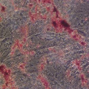
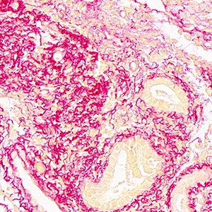
Using an immunohistochemical technique, we have studied the distribution of kynuneric acid (KYNA) and 3-hydroxyanthranilic acid (3-HAA) in a rat brain injury model (trauma). The study was carried out inducing a cerebral ablation of the frontal motor cortex. Two mouse monoclonal specific antibodies previously developed by our group directed against KYNA and 3-HAA were used. In control animals (sham-operated), the expression of both KYNA and 3-HAA was not observed. In animals in which the ablation was performed, the highest number of immunoreactive cells containing KYNA or 3-HAA was observed in the region surrounding the lesion and the number of these cells decreased moving away from the lesion. KYNA and 3-HAA were also observed in the white matter (ipsilateral side) located close to the injured region and in some cells placed in the white matter of the contralateral side. The distribution of KYNA and 3-HAA perfectly matched with the peripheral injured regions. The results found were identical independently of the perfusion date of animals (17, 30 or 54 days after brain injury). For the first time, the presence of KYNA and 3-HAA has been described in a rat trauma model. Moreover, by using a double immunocytochemistry protocol, it has been demonstrated that both metabolites were located in astrocytes. The findings observed suggest that, in cerebral trauma, KYNA and 3-HAA are involved in tissue damage and that these compounds could act, respectively, as a neuroprotector and a neurotoxic. This means that, in trauma, a counterbalance occurs and that a regulation of the indoleamine 2,3 dioxygenase (IDO) pathway could be required after a brain injury in order to decrease the deleterious effects of ending metabolites (the neurotoxic picolinic acid). Moreover, the localization of KYNA and 3-HAA in the contralateral side of the lesion suggests that the IDO pathway is also involved in the sprouting and pathfinding that follows a traumatic brain injury.
Downloads
Publication Facts
Reviewer profiles N/A
Author statements
- Editor & editorial board
- profiles
- Academic society
- N/A
- Publisher
- PAGEPress Publications, Pavia, Italy

To learn about these publication facts, click ![]()
PF is maintained by the Public Knowledge Project
Citations


Supporting Agencies
Gemacbio S.A. Laboratories (Saint Jean d’Illac, France), IDRPHT (Talence, France)How to Cite
PAGEPress has chosen to apply the Creative Commons Attribution NonCommercial 4.0 International License (CC BY-NC 4.0) to all manuscripts to be published.
Similar Articles
- Arturo Mangas, Javier Yajeya, Noelia González, Isabel Ruiz, Marianny Pernía, Michel Geffard, Rafael Coveñas, Gemst: a taylor-made combination that reverts neuroanatomical changes in stroke , European Journal of Histochemistry: Vol. 61 No. 2 (2017)
- E. Akat, H. Arıkan, B. Göçmen, Histochemical and biometric study of the gastrointestinal system of Hyla orientalis (Bedriaga, 1890) (Anura, Hylidae) , European Journal of Histochemistry: Vol. 58 No. 4 (2014)
- Yankun Wang, Yuning Liu, Yawei Wang, Ao Zhang, Wenqian Xie, Haolin Zhang, Qiang Weng, Meiyu Xu, Investigation of seasonal changes in lipid synthesis and metabolism-related genes in the oviduct of Chinese brown frog (Rana dybowskii) , European Journal of Histochemistry: Vol. 67 No. 4 (2023)
- M. Lehmann, F. Martin, K. Mannigel, K. Kaltschmidt, U. Sack, U. Anderer, Three-dimensional scaffold-free fusion culture: the way to enhance chondrogenesis of in vitro propagated human articular chondrocytes , European Journal of Histochemistry: Vol. 57 No. 4 (2013)
- A. Mangas, J. Yajeya, N. González, I. Ruiz, M. Geffard, R. Coveñas, 3-hydroxi-anthranilic acid is early expressed in stroke , European Journal of Histochemistry: Vol. 60 No. 4 (2016)
- M. J. Galvão, A. Santos, M. D. Ribeiro, A. Ferreira, F. Nolasco, Optimization of the tartrate-resistant acid phosphatase detection by histochemical method , European Journal of Histochemistry: Vol. 55 No. 1 (2011)
- D. Ami, M. Di Segni, M. Forcella, V. Meraviglia, M. Baccarin, S.M. Doglia, G. Terzoli, Role of water in chromosome spreading and swelling induced by acetic acid treatment: a FTIR spectroscopy study , European Journal of Histochemistry: Vol. 58 No. 1 (2014)
- L. Vinci, A. Ravarino, V. Fanos, A.G. Naccarato, G. Senes, C. Gerosa, G. Bevilacqua, G. Faa, R. Ambu, Immunohistochemical markers of neural progenitor cells in the early embryonic human cerebral cortex , European Journal of Histochemistry: Vol. 60 No. 1 (2016)
- S. Strobel, J.A. Encarnação, N.I. Becker, T.E. Trenczek, Histological and histochemical analysis of the gastrointestinal tract of the common pipistrelle bat (Pipistrellus pipistrellus) , European Journal of Histochemistry: Vol. 59 No. 2 (2015)
- I.M.S. Paulsen, H. Dimke, S. Frische, A single simple procedure for dewaxing, hydration and heat-induced epitope retrieval (HIER) for immunohistochemistry in formalin fixed paraffin-embedded tissue , European Journal of Histochemistry: Vol. 59 No. 4 (2015)
You may also start an advanced similarity search for this article.