Peribiliary gland damage due to liver transplantation involves peribiliary vascular plexus and vascular endothelial growth factor
Accepted: 23 April 2019
Supplementary: 0
HTML: 8
All claims expressed in this article are solely those of the authors and do not necessarily represent those of their affiliated organizations, or those of the publisher, the editors and the reviewers. Any product that may be evaluated in this article or claim that may be made by its manufacturer is not guaranteed or endorsed by the publisher.
Authors
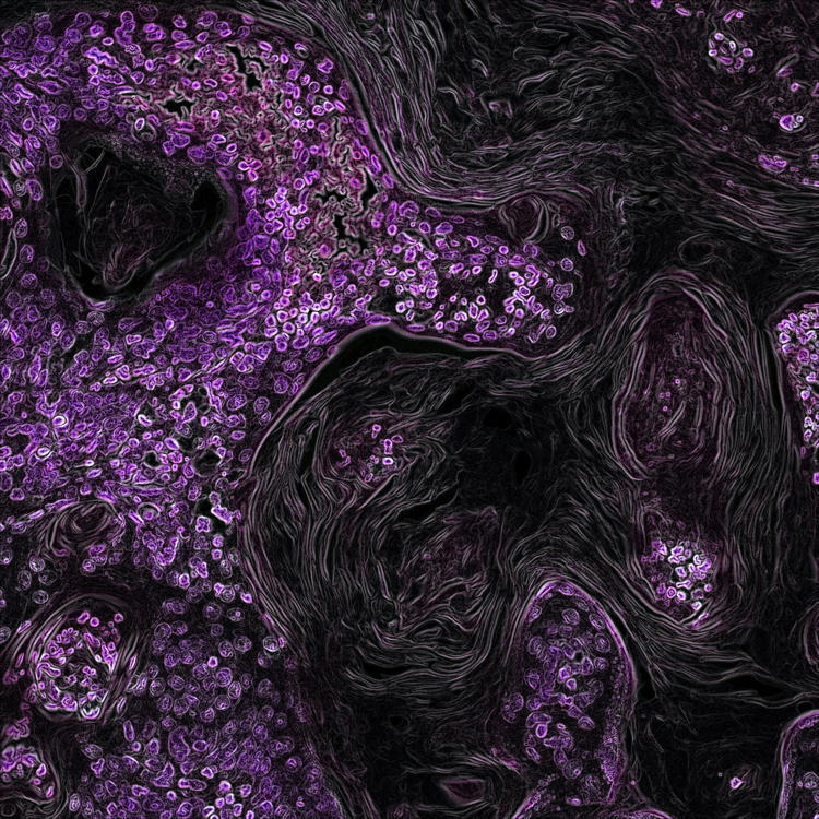
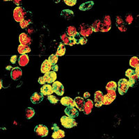
Extrahepatic bile ducts are characterized by the presence of peribiliary glands (PBGs), which represent stem cell niches implicated in biliary regeneration. Orthotopic liver transplantation may be complicated by non-anastomotic strictures (NAS) of the bile ducts, which have been associated with ischemic injury of PBGs and occur more frequently in livers obtained from donors after circulatory death than in those from brain-dead donors. The aims of the present study were to investigate the PBG phenotype in bile ducts after transplantation, the integrity of the peribiliary vascular plexus (PVP) around PBGs, and the expression of vascular endothelial growth factor-A (VEGF-A) by PBGs. Transplanted ducts obtained from patients who underwent liver transplantation were studied (N=62). Controls included explanted bile duct samples not used for transplantation (N=10) with normal histology. Samples were processed for histology, immunohistochemistry and immunofluorescence. Surface epithelium is severely injured in transplanted ducts; PBGs are diffusely damaged, particularly in ducts obtained from circulatory-dead compared to brain-dead donors. PVP is reduced in transplanted compared to controls. PBGs in transplanted ducts contain more numerous progenitor and proliferating cells compared to controls, show higher positivity for VEGF-A compared to controls, and express VEGF receptor-2. In conclusion, PBGs and associated PVP are damaged in transplanted extrahepatic bile ducts; however, an activation of the PBG niche takes place and is characterized by proliferation and VEGF-A expression. This response could have a relevant role in reconstituting biliary epithelium and vascular plexus and could be implicated in the genesis of non-anastomotic strictures.


Supporting Agencies
Sapienza University of Rome, European Society for Organ Transplantation, Vesta Therapeutics (Bethesda, MD, USA)How to Cite
PAGEPress has chosen to apply the Creative Commons Attribution NonCommercial 4.0 International License (CC BY-NC 4.0) to all manuscripts to be published.
Similar Articles
- E. Tarantola, V. Bertone, G. Milanesi, E. Capelli, A. Ferrigno, D. Neri, M. Vairetti, S. Barni, I. Freitas, Dipeptidylpeptidase-ÂIV, a key enzyme for the degradation of incretins and neuropeptides: activity and expression in the liver of lean and obese rats , European Journal of Histochemistry: Vol. 56 No. 4 (2012)
- V. Bertone, E. Tarantola, A. Ferrigno, E. Gringeri, S. Barni, M. Vairetti, I. Freitas, Altered alkaline phosphatase activity in obese Zucker rats liver respect to lean Zucker and Wistar rats discussed in terms of all putative roles ascribed to the enzyme , European Journal of Histochemistry: Vol. 55 No. 1 (2011)
- Matias Garrido, Camila Escobar, Constanza Zamora, Carolina Rejas, Juan Varas, Mario Párraga, Sebastián San Martin, Sandra Montedonico, Bile duct ligature in young rats: A revisited animal model for biliary atresia , European Journal of Histochemistry: Vol. 61 No. 3 (2017)
- E. Tarantola, V. Bertone, G. Milanesi, C. Gruppi, A. Ferrigno, M. Vairetti, S. Barni, I. Freitas, Dipeptidylpeptidase-IV activity and expression reveal decreased damage to the intrahepatic biliary tree in fatty livers submitted to subnormothermic machine-perfusion respect to conventional cold storage , European Journal of Histochemistry: Vol. 58 No. 3 (2014)
- Simone Carotti, Giuseppe Perrone, Michelina Amato, Umberto Vespasiani Gentilucci, Daniela Righi, Maria Francesconi, Claudio Pellegrini, Francesca Zalfa, Maria Zingariello, Antonio Picardi, Andrea Onetti Muda, Sergio Morini, Reelin expression in human liver of patients with chronic hepatitis C infection , European Journal of Histochemistry: Vol. 61 No. 1 (2017)
- Claudia Frick, Hanna Luisa Martin, Johanna Bruder, Kerstin Lang, Heinz Breer, Topographic distribution pattern of morphologically different G cells in the murine antral mucosa , European Journal of Histochemistry: Vol. 61 No. 3 (2017)
- Tadashi Yasui, Kenya Miyata, Chie Nakatsuka, Azuma Tsukise, Hiroshi Gomi, Morphological and histochemical characterization of the secretory epithelium in the canine lacrimal gland , European Journal of Histochemistry: Vol. 65 No. 4 (2021)
- Xilin Ge, Caoxin Huang, Wenting Chen, Chen Yang, Wenfang Huang, Jia Li, Shuyu Yang, Effect of Danggui Buxue decoction on hypoxia-induced injury of retinal Müller cells in vitro , European Journal of Histochemistry: Vol. 68 No. 4 (2024)
- S. Strobel, J.A. Encarnação, N.I. Becker, T.E. Trenczek, Histological and histochemical analysis of the gastrointestinal tract of the common pipistrelle bat (Pipistrellus pipistrellus) , European Journal of Histochemistry: Vol. 59 No. 2 (2015)
- S. Huang, S. Guo, F. Guo, Q. Yang, X. Xiao, M. Murata, S. Ohnishi, S. Kawanishi, N. Ma, CD44v6 expression in human skin keratinocytes as a possible mechanism for carcinogenesis associated with chronic arsenic exposure , European Journal of Histochemistry: Vol. 57 No. 1 (2013)
You may also start an advanced similarity search for this article.
Publication Facts
Reviewer profiles N/A
Author statements
- Editor & editorial board
- profiles
- Academic society
- N/A
- Publisher
- PAGEPress Publications, Pavia, Italy

To learn about these publication facts, click ![]()
PF is maintained by the Public Knowledge Project