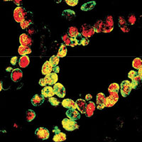
Peribiliary gland damage due to liver transplantation involves peribiliary vascular plexus and vascular endothelial growth factor

Submitted: 7 March 2019
Accepted: 23 April 2019
Published: 10 May 2019
Accepted: 23 April 2019
Abstract Views: 1554
PDF: 618
Supplementary: 0
HTML: 8
Supplementary: 0
HTML: 8
Publisher's note
All claims expressed in this article are solely those of the authors and do not necessarily represent those of their affiliated organizations, or those of the publisher, the editors and the reviewers. Any product that may be evaluated in this article or claim that may be made by its manufacturer is not guaranteed or endorsed by the publisher.
All claims expressed in this article are solely those of the authors and do not necessarily represent those of their affiliated organizations, or those of the publisher, the editors and the reviewers. Any product that may be evaluated in this article or claim that may be made by its manufacturer is not guaranteed or endorsed by the publisher.
Similar Articles
- N. Tolosa de Talamoni, A. Pérez, R. Riis, C. Smith, M. L. Norman, R. H. Wasserman, Comparative immunolocalization of the plasma membrane calcium pump and calbindin D28K in chicken retina during embryonic development , European Journal of Histochemistry: Vol. 46 No. 4 (2002)
- I.M.S. Paulsen, H. Dimke, S. Frische, A single simple procedure for dewaxing, hydration and heat-induced epitope retrieval (HIER) for immunohistochemistry in formalin fixed paraffin-embedded tissue , European Journal of Histochemistry: Vol. 59 No. 4 (2015)
- NACS Wong, M Herriot, F Rae, An immunohistochemical study and review of potential markers of human intestinal M cells , European Journal of Histochemistry: Vol. 47 No. 2 (2003)
- H. Huang, W. Wang, P. Liu, Y. Jiang, Y. Zhao, H. Wei, W. Niu, TRPC1 expression and distribution in rat hearts , European Journal of Histochemistry: Vol. 53 No. 4 (2009)
- E Redondo, A Franco, AJ Masot, S Regodón, Ultrastructural and immunocytochemical characterization of interstitial cells in pre- and postnatal developing sheep pineal gland , European Journal of Histochemistry: Vol. 45 No. 3 (2001)
- S Passinen, T Ylikomi, Evidence for the existence of an oligomeric, non-DNA-binding complex of the progesterone receptor in the cytoplasm , European Journal of Histochemistry: Vol. 47 No. 3 (2003)
- I Kasacka, AM Humenczyk-Zybala, M Niczyporuk, G Mycko, Morphometric evaluation of murine pulmonary mast cells in experimental hemorrhagic shock , European Journal of Histochemistry: Vol. 48 No. 2 (2004)
- C Shi, G Zhou, Y Zhu, Y Su, T Cheng, HE Zhau, LWK Chung, Quantum dots-based multiplexed immunohistochemistry of protein expression in human prostate cancer cells , European Journal of Histochemistry: Vol. 52 No. 2 (2008)
- C Campanella, F Bucchieri, NM Ardizzone, A Marino Gammazza, A Montalbano, A Ribbene, V Di Felice, M Bellafiore, S David, F Rappa, M Marasà , G Peri, F Farina, AM Czarnecka, E Conway de Macario, AJL Macario, G Zummo, F Cappello, Upon oxidative stress, the antiapoptotic Hsp60/procaspase-3 complex persists in mucoepidermoid carcinoma cells , European Journal of Histochemistry: Vol. 52 No. 4 (2008)
- A Sedo, R MalÃk, K Drbal, V Lisá, K Vlasicová, V Mares, Dipeptidyl peptidase IV in two human glioma cell lines , European Journal of Histochemistry: Vol. 45 No. 1 (2001)
<< < 51 52 53 54 55 56 57 58 59 60 > >>
You may also start an advanced similarity search for this article.

 https://doi.org/10.4081/ejh.2019.3022


