
A method for semi-automated image analysis of HLA class I tumour epithelium expression in rectal cancer
All claims expressed in this article are solely those of the authors and do not necessarily represent those of their affiliated organizations, or those of the publisher, the editors and the reviewers. Any product that may be evaluated in this article or claim that may be made by its manufacturer is not guaranteed or endorsed by the publisher.
Accepted: 20 April 2019
Authors
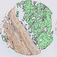
Biomarkers may hold the key towards development and improvement of personalized cancer treatment. For instance, tumour expression of immune system-related proteins may reveal the tumour immune status and, accordingly, determine choice for type of immunotherapy. Therefore, objective evaluation of tumour biomarker expression is needed but often challenging. For instance, human leukocyte antigen (HLA) class I tumour epithelium expression is cumbersome to quantify by eye due to its presence on both tumour epithelial cells and tumour stromal cells, as well as tumour-infiltrating immune cells. In this study, we solved this problem by setting up an immunohistochemical (IHC) double staining using a tissue microarray (TMA) of rectal tumours wherein HLA class I expression was coloured with a blue chromogen, whereas non-epithelial tissue was visualized with a brown chromogen. We subsequently developed a semi-automated image analysis method that identified tumour epithelium as well as the percentage of HLA class I-positive tumour epithelium. Using this technique, we compared HCA2/HC10 and EMR8-5 antibodies for the assessment of HLA class I tumour expression and concluded that EMR8-5 is the superior antibody for this purpose. This IHC double staining can in principle be used for scoring of any biomarker expressed by tumour epithelium.
Downloads
Citations


