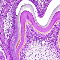
Expression, localization and synthesis of small leucine-rich proteoglycans in developing mouse molar tooth germ

Submitted: 2 December 2019
Accepted: 31 January 2020
Published: 10 February 2020
Accepted: 31 January 2020
Abstract Views: 1197
PDF: 882
HTML: 5
HTML: 5
Publisher's note
All claims expressed in this article are solely those of the authors and do not necessarily represent those of their affiliated organizations, or those of the publisher, the editors and the reviewers. Any product that may be evaluated in this article or claim that may be made by its manufacturer is not guaranteed or endorsed by the publisher.
All claims expressed in this article are solely those of the authors and do not necessarily represent those of their affiliated organizations, or those of the publisher, the editors and the reviewers. Any product that may be evaluated in this article or claim that may be made by its manufacturer is not guaranteed or endorsed by the publisher.
Similar Articles
- S Montagnani, L Postiglione, G Giordano-Lanza, F Di Meglio, C Castaldo, S Sciorio, N Montuori, G Di Spigna, P Ladogana, A Oriente, G Rossi, Granulocyte macrophage colony stimulating factor (GM-CSF) biological actions on human dermal fibroblasts , European Journal of Histochemistry: Vol. 45 No. 3 (2001)
- E. Fantinato, L. Milani, G. Sironi, Sox9 expression in canine epithelial skin tumors , European Journal of Histochemistry: Vol. 59 No. 3 (2015)
- H. Zhang, P. Yu, S. Zhong, T. Ge, S. Peng, Z. Zhou, X. Guo, Gliocyte and synapse analyses in cerebral ganglia of the Chinese mitten crab, Eriocheir sinensis: ultrastructural study , European Journal of Histochemistry: Vol. 60 No. 3 (2016)
- Yiming Zhang, Wanqiong Zheng, Liang Zhang, Yechun Gu, Lihe Zhu, Yingpeng Huang, LncRNA FBXO18-AS promotes gastric cancer progression by TGF-β1/Smad signaling , European Journal of Histochemistry: Vol. 67 No. 2 (2023)
- Arianna Casini, Giorgio Vivacqua, Rosa Vaccaro, Anastasia Renzi, Stefano Leone, Luigi Pannarale, Antonio Franchitto, Paolo Onori, Romina Mancinelli, Eugenio Gaudio, Expression and role of cocaine-amphetamine regulated transcript (CART) in the proliferation of biliary epithelium , European Journal of Histochemistry: Vol. 67 No. 4 (2023)
- M.G. Manfredi Romanini, Postgenomic histochemistry , European Journal of Histochemistry: Vol. 47 No. 1 (2003)
- S Fakan, Ultrastructural cytochemical analyses of nuclear functional architecture , European Journal of Histochemistry: Vol. 48 No. 1 (2004)
- B Bilinska, M Kotula-Balak, J Sadowska, Morphology and function of human Leydig cells in vitro. Immunocytochemical and radioimmunological analyses , European Journal of Histochemistry: Vol. 53 No. 1 (2009)
- G D'Ancona Lunetta, ML Michelucci, Engagement of the periesophageal ring during Holothuria polii response to erythrocyte injection , European Journal of Histochemistry: Vol. 46 No. 2 (2002)
- A.M. Schläfli, S. Berezowska, O. Adams, R. Langer, M.P. Tschan, Reliable LC3 and p62 autophagy marker detection in formalin fixed paraffin embedded human tissue by immunohistochemistry , European Journal of Histochemistry: Vol. 59 No. 2 (2015)
<< < 41 42 43 44 45 46 47 48 > >>
You may also start an advanced similarity search for this article.

 https://doi.org/10.4081/ejh.2020.3092


