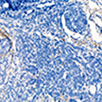
Aberrant expression of CCDC69 in breast cancer and its clinicopathologic significance

Submitted: 10 December 2020
Accepted: 27 January 2021
Published: 22 February 2021
Abstract Views: 931
PDF: 605
HTML: 19
Publisher's note
All claims expressed in this article are solely those of the authors and do not necessarily represent those of their affiliated organizations, or those of the publisher, the editors and the reviewers. Any product that may be evaluated in this article or claim that may be made by its manufacturer is not guaranteed or endorsed by the publisher.

Authors

Coiled-coil domain-containing protein 69 (CCDC69) is a novel gene and limited knowledge in known in breast cancer. In the present study, we aimed to explore the relationship between CCDC69 and breast cancer, demonstrate the clinicopathological significance and prognostic role of CCDC69 in breast cancer, and analyze the possible mechanism of CCDC69 affecting the prognosis of breast cancer. First, from GEO database, TIMER, GEPIA, and OncoLnc, we select CCDC69 as the potential gene which closely involved in breast cancer progression. Next, by real-time PCR detection, the expression of CCDC69 in breast cancer tissue was notably lower than that in normal breast tissues (p=0.0002). In addition, our immunohistochemistry (IHC) indicated that the positive expression rate of CCDC69 in the triple-negative breast cancer (TNBC) was lower than that in the non-TNBC (p=0.0362), and it was negatively correlated with the expression of Ki67 (p=0.001). Further enrichment analysis of CCDC69 and the similar genes performed on FunRich3.1.3 revealed that these genes were significantly associated with fat differentiation, and most of them were related to peroxisome proliferator-activated receptor (PPAR) signal pathway. Collectively, our findings suggest that CCDC69 is down regulated in breast cancer tissue especially in TNBC which has higher malignant grade and poorer clinical prognosis.

Dimensions

Altmetric

PlumX Metrics

Downloads

Download data is not yet available.

Citations

Bray F, Ferlay J, Soerjomataram I, Siegel RL, Torre LA, Jemal A. Global cancer statistics 2018: GLOBOCAN estimates of incidence and mortality worldwide for 36 cancers in 185 countries. CA Cancer J Clin 2018;68:394-424. DOI: https://doi.org/10.3322/caac.21492
Hwang ES, Hyslop T, Hendrix LH, Duong S, Bedrosian I, Price E, et al. Phase II single-arm study of preoperative letrozole for estrogen receptor-positive postmenopausal ductal carcinoma in situ: CALGB 40903 (alliance). J Clin Oncol 2020;38:JCO1900510. DOI: https://doi.org/10.1200/JCO.19.00510
Mavroudis D, Saloustros E, Malamos N, Kakolyris S, Boukovinas I, Papakotoulas P, et al. Corrigendum to Six versus 12 months of adjuvant trastuzumab in combination with dose-dense chemotherapy for women with HER2-positive breast cancer: a multicenter randomized study by the Hellenic Oncology Research Group (HORG): Annals of Oncology, Volume 26, Issue 7, July 2015, Pages 1333-1340. Ann Oncol 2020;31:444-5. DOI: https://doi.org/10.1016/j.annonc.2020.01.004
Kurian AW, Ward KC, Abrahamse P, Hamilton AS, Deapen D, Morrow M, et al. Association of germline genetic testing results with locoregional and systemic therapy in patients with breast cancer. JAMA Oncol 2020;6:e196400. DOI: https://doi.org/10.1001/jamaoncol.2019.6400
Vasan N, Toska E, Scaltriti M. Overview of the relevance of PI3K pathway in HR-positive breast cancer. Ann Oncol 2019;30:x3-x11. DOI: https://doi.org/10.1093/annonc/mdz281
Schmid P, Abraham J, Chan S, Wheatley D, Brunt AM, Nemsadze G, et al. Capivasertib plus paclitaxel versus placebo plus paclitaxel as first-line therapy for metastatic triple-negative breast cancer: The PAKT Trial. J Clin Oncol 2020;38:423-33. DOI: https://doi.org/10.1200/JCO.19.00368
Baxter JS, Leavy OC, Dryden NH, Maguire S, Johnson N, Fedele V, et al. Capture Hi-C identifies putative target genes at 33 breast cancer risk loci. Nat Commun 2018;9:1028. DOI: https://doi.org/10.1038/s41467-018-03411-9
Zhou J, Lei J, Wang J, Lian CL, Hua L, He ZY, et al. Bioinformatics-based discovery of CKLF-Like MARVEL transmembrane member 5 as a novel biomarker for breast cancer. Front Cell Dev Biol 2019;7:361. DOI: https://doi.org/10.3389/fcell.2019.00361
Li Y, Umbach DM, Bingham A, Li QJ, Zhuang Y, Li L. Putative biomarkers for predicting tumor sample purity based on gene expression data. BMC Genomics 2019;20:1021. DOI: https://doi.org/10.1186/s12864-019-6412-8
Yoshihara K, Shahmoradgoli M, Martinez E, Vegesna R, Kim H, Torres-Garcia W, et al. Inferring tumour purity and stromal and immune cell admixture from expression data. Nat Commun 2013;4:2612. DOI: https://doi.org/10.1038/ncomms3612
Cui L, Zhou F, Chen C, Wang CC. Overexpression of CCDC69 activates p14(ARF)/MDM2/p53 pathway and confers cisplatin sensitivity. J Ovarian Res 2019;12:4. DOI: https://doi.org/10.1186/s13048-019-0479-3
Pal D, Wu D, Haruta A, Matsumura F, Wei Q. Role of a novel coiled-coil domain-containing protein CCDC69 in regulating central spindle assembly. Cell Cycle 2010;9:4117-29. DOI: https://doi.org/10.4161/cc.9.20.13387
Lesniewski LA, Hosch SE, Neels JG, de Luca C, Pashmforoush M, Lumeng CN, et al. Bone marrow-specific Cap gene deletion protects against high-fat diet-induced insulin resistance. Nat Med 2007;13:455-62. DOI: https://doi.org/10.1038/nm1550
Ribon V, Printen JA, Hoffman NG, Kay BK, Saltiel AR. A novel, multifuntional c-Cbl binding protein in insulin receptor signaling in 3T3-L1 adipocytes. Mol Cell Biol 1998;18:872-9. DOI: https://doi.org/10.1128/MCB.18.2.872
Chen YZ, Xue JY, Chen CM, Yang BL, Xu QH, Wu F, et al. PPAR signaling pathway may be an important predictor of breast cancer response to neoadjuvant chemotherapy. Cancer Chemother Pharmacol 2012;70:637-44. DOI: https://doi.org/10.1007/s00280-012-1949-0
Nimmrich I, Sieuwerts AM, Meijer-van Gelder ME, Schwope I, Bolt-de Vries J, Harbeck N, et al. DNA hypermethylation of PITX2 is a marker of poor prognosis in untreated lymph node-negative hormone receptor-positive breast cancer patients. Breast Cancer Res Treat 2008;111:429-37. DOI: https://doi.org/10.1007/s10549-007-9800-8
Gao C, Zhuang J, Li H, Liu C, Zhou C, Liu L, et al. Development of a risk scoring system for evaluating the prognosis of patients with Her2-positive breast cancer. Cancer Cell Int 2020;20:121. DOI: https://doi.org/10.1186/s12935-020-01175-1
Penault-Llorca F, Radosevic-Robin N. Ki67 assessment in breast cancer: an update. Pathology 2017;49:166-71. DOI: https://doi.org/10.1016/j.pathol.2016.11.006
Jurikova M, Danihel L, Polak S, Varga I. Ki67, PCNA, and MCM proteins: Markers of proliferation in the diagnosis of breast cancer. Acta Histochem 2016;118:544-52. DOI: https://doi.org/10.1016/j.acthis.2016.05.002
Regan MM, Pagani O, Francis PA, Fleming GF, Walley BA, Kammler R, et al. Predictive value and clinical utility of centrally assessed ER, PgR, and Ki-67 to select adjuvant endocrine therapy for premenopausal women with hormone receptor-positive, HER2-negative early breast cancer: TEXT and SOFT trials. Breast Cancer Res Treat 2015;154:275-86. DOI: https://doi.org/10.1007/s10549-015-3612-z
Cabrera-Galeana P, Munoz-Montano W, Lara-Medina F, Alvarado-Miranda A, Perez-Sanchez V, Villarreal-Garza C, et al. Ki67 changes identify worse outcomes in residual breast cancer tumors after neoadjuvant chemotherapy. Oncologist 2018;23:670-8. DOI: https://doi.org/10.1634/theoncologist.2017-0396
Sueta A, Yamamoto Y, Hayashi M, Yamamoto S, Inao T, Ibusuki M, et al. Clinical significance of pretherapeutic Ki67 as a predictive parameter for response to neoadjuvant chemotherapy in breast cancer: is it equally useful across tumor subtypes? Surgery 2014;155:927-35. DOI: https://doi.org/10.1016/j.surg.2014.01.009
Steward L, Conant L, Gao F, Margenthaler JA. Predictive factors and patterns of recurrence in patients with triple negative breast cancer. Ann Surg Oncol 2014;21:2165-71. DOI: https://doi.org/10.1245/s10434-014-3546-4
Navratil J, Fabian P, Palacova M, Petrakova K, Vyzula R, Svoboda M. [Triple negative breast cancer].[Article in Czech]. Klin Onkol 2015;28:405-15.
da Silva JL, Cardoso Nunes NC, Izetti P, de Mesquita GG, de Melo AC. Triple negative breast cancer: A thorough review of biomarkers. Crit Rev Oncol Hematol 2020;145:102855. DOI: https://doi.org/10.1016/j.critrevonc.2019.102855
Mendes TF, Kluskens LD, Rodrigues LR. Triple negative breast cancer: Nanosolutions for a big challenge. Adv Sci (Weinh) 2015;2:1500053. DOI: https://doi.org/10.1002/advs.201500053
Li X, Yang J, Peng L, Sahin AA, Huo L, Ward KC, et al. Triple-negative breast cancer has worse overall survival and cause-specific survival than non-triple-negative breast cancer. Breast Cancer Res Treat 2017;161:279-87. DOI: https://doi.org/10.1007/s10549-016-4059-6
Wang Y, Xu H, Sun G, Xue M, Sun S, Huang T, et al. Transcriptome analysis of the effects of fasting caecotrophy on hepatic lipid metabolism in New Zealand rabbits. Animals (Basel) 2019;9:648. DOI: https://doi.org/10.3390/ani9090648
Sultan G, Zubair S, Tayubi IA, Dahms HU, Madar IH. Towards the early detection of ductal carcinoma (a common type of breast cancer) using biomarkers linked to the PPAR(gamma) signaling pathway. Bioinformation 2019;15:799-805. DOI: https://doi.org/10.6026/97320630015799
Joseph C, Papadaki A, Althobiti M, Alsaleem M, Aleskandarany MA, Rakha EA. Breast cancer intratumour heterogeneity: current status and clinical implications. Histopathology 2018;73:717-31. DOI: https://doi.org/10.1111/his.13642
Van Keymeulen A, Lee MY, Ousset M, Brohee S, Rorive S, Giraddi RR, et al. Reactivation of multipotency by oncogenic PIK3CA induces breast tumour heterogeneity. Nature 2015;525:119-23. DOI: https://doi.org/10.1038/nature14665
Scheel C, Weinberg RA. Cancer stem cells and epithelial-mesenchymal transition: concepts and molecular links. Semin Cancer Biol 2012;22:396-403. DOI: https://doi.org/10.1016/j.semcancer.2012.04.001
Tsilimigras DI, Oikonomou EK, Moris D, Schizas D, Economopoulos KP, Mylonas KS. Stem cell therapy for congenital heart disease: A systematic review. Circulation 2017;136:2373-85. DOI: https://doi.org/10.1161/CIRCULATIONAHA.117.029607
Issemann I, Green S. Activation of a member of the steroid hormone receptor superfamily by peroxisome proliferators. Nature 1990;347:645-50. DOI: https://doi.org/10.1038/347645a0
Polvani S, Tarocchi M, Tempesti S, Bencini L, Galli A. Peroxisome proliferator activated receptors at the crossroad of obesity, diabetes, and pancreatic cancer. World J Gastroenterol 2016;22:2441-59. DOI: https://doi.org/10.3748/wjg.v22.i8.2441
Zhang Y, Zhang X, Wang J, Shen Y, Tang X, Yu F, et al. Expression and function of PPARs in Cancer stem cells. Curr Stem Cell Res Ther 2016;11:226-34. DOI: https://doi.org/10.2174/1574888X10666150728122921
Ishay-Ronen D, Diepenbruck M, Kalathur RKR, Sugiyama N, Tiede S, Ivanek R, et al. Gain fat-lose metastasis: Converting invasive breast cancer cells into adipocytes inhibits cancer metastasis. Cancer Cell 2019;35:17-32.e6. DOI: https://doi.org/10.1016/j.ccell.2018.12.002
Ishay-Ronen D, Christofori G. Targeting cancer cell metastasis by converting cancer cells into fat. Cancer Res 2019;79:5471-5. DOI: https://doi.org/10.1158/0008-5472.CAN-19-1242
Mueller E, Sarraf P, Tontonoz P, Evans RM, Martin KJ, Zhang M, et al. Terminal differentiation of human breast cancer through PPAR gamma. Mol Cell 1998;1:465-70. DOI: https://doi.org/10.1016/S1097-2765(00)80047-7
Zheng ZH, Yang Y, Lu XH, Zhang H, Shui XX, Liu C, et al. Mycophenolic acid induces adipocyte-like differentiation and reversal of malignancy of breast cancer cells partly through PPARgamma. Eur J Pharmacol 2011;658:1-8. DOI: https://doi.org/10.1016/j.ejphar.2011.01.068
Wu L, Wang K, Wang W, Wen Z, Wang P, Liu L, et al. Glucagon-like peptide-1 ameliorates cardiac lipotoxicity in diabetic cardiomyopathy via the PPARalpha pathway. Aging Cell 2018;17:e12763. DOI: https://doi.org/10.1111/acel.12763
Akune T, Ohba S, Kamekura S, Yamaguchi M, Chung UI, Kubota N, et al. PPARgamma insufficiency enhances osteogenesis through osteoblast formation from bone marrow progenitors. J Clin Invest 2004;113:846-55. DOI: https://doi.org/10.1172/JCI200419900
Osinski V, Bauknight DK, Dasa SSK, Harms MJ, Kroon T, Marshall MA, et al. In vivo liposomal delivery of PPARalpha/gamma dual agonist tesaglitazar in a model of obesity enriches macrophage targeting and limits liver and kidney drug effects. Theranostics 2020;10:585-601. DOI: https://doi.org/10.7150/thno.36572
Davalos-Salas M, Montgomery MK, Reehorst CM, Nightingale R, Ng I, Anderton H, et al. Deletion of intestinal Hdac3 remodels the lipidome of enterocytes and protects mice from diet-induced obesity. Nat Commun 2019;10:5291. DOI: https://doi.org/10.1038/s41467-019-13180-8
Yu W, Li D, Zhang Y, Li C, Zhang C, Wang L. MiR-142-5p acts as a significant regulator through promoting proliferation, invasion, and migration in breast cancer modulated by targeting SORBS1. Technol Cancer Res Treat 2019;18:1533033819892264. DOI: https://doi.org/10.1177/1533033819892264
Song L, Chang R, Dai C, Wu Y, Guo J, Qi M, et al. SORBS1 suppresses tumor metastasis and improves the sensitivity of cancer to chemotherapy drug. Oncotarget 2017;8:9108-22. DOI: https://doi.org/10.18632/oncotarget.12851

How to Cite

Li, J., Zhang, P., & Xia, Y. (2021). Aberrant expression of <em>CCDC69</em> in breast cancer and its clinicopathologic significance. European Journal of Histochemistry, 65(1). https://doi.org/10.4081/ejh.2021.3207