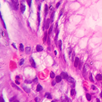
Efficacy of immunohistochemical staining in detecting Helicobacter pylori in Saudi patients with minimal and atypical infection

Submitted: 8 February 2021
Accepted: 5 May 2021
Published: 20 July 2021
Abstract Views: 1154
PDF: 536
HTML: 117
Publisher's note
All claims expressed in this article are solely those of the authors and do not necessarily represent those of their affiliated organizations, or those of the publisher, the editors and the reviewers. Any product that may be evaluated in this article or claim that may be made by its manufacturer is not guaranteed or endorsed by the publisher.

Authors

Gastric Helicobacter pylori infection is diagnosed based on histopathological evaluation of gastric mucosal biopsies, urease test, urea breath test, H. pylori culturing, or direct detection using polymerase chain reaction (PCR). This study aimed to evaluate the efficacy of immunohistochemical (IHC) staining in detecting H. pylori in gastric biopsies from patients with chronic gastritis and minimal or atypical infection. Gastric biopsies from 50 patients with chronic gastritis were subjected to routine haematoxylin and eosin (H-E), modified Giemsa, and IHC staining. The results of staining were compared with those of quantitative real-time PCR (qRT-PCR). The qRT-PCR analysis identified 32 (64%) H. pylori-positive cases, whereas IHC, H-E, and modified Giemsa staining identified 29 (58%), 27 (54%), and 21 (42%) positive cases. The sensitivity of IHC staining (87.50%) was higher than that of H-E (59.38%) and modified Giemsa (43.75%) staining. The specificity of H-E, modified Giemsa, and IHC staining was 55.56%, 61.11%, and 94.44%, respectively. IHC staining exhibited the highest diagnostic accuracy (90%), followed by H-E (58%) and modified Giemsa (50%) staining. Active gastritis, intestinal metaplasia, and lymphoid follicles were detected in 32 (64%), 4 (8%), and 22 (44%) cases, respectively, and all of these cases were H. pylori positive. In contrast to routine H-E and modified Giemsa staining, IHC allows for the accurate H. pylori detection in cases with minimal or atypical infection. Moreover, IHC can be an alternative diagnostic method to qRT-PCR for detection of H. pylori in such cases.

Dimensions

Altmetric

PlumX Metrics

Downloads

Download data is not yet available.

Citations

Shimizu T, Akamatsu T, Ota H, Katsuyama T. Immunohistochemical detection of Helicobacter pylori in the surface mucous gel layer and its clinicopathological significance. Helicobacter 1996;1:197-206. DOI: https://doi.org/10.1111/j.1523-5378.1996.tb00040.x
Garza-Gonzalez E, Perez-Perez GI, Maldonado-Garza HJ, Bosques-Padilla FJ. A review of Helicobacter pylori diagnosis, treatment, and methods to detect eradication. World J Gastroenterol 2014;20:1438-49. DOI: https://doi.org/10.3748/wjg.v20.i6.1438
Megraud F, Lehours P. Helicobacter pylori detection and antimicrobial susceptibility testing. Clin Microbiol Rev 2007;20:280-322. DOI: https://doi.org/10.1128/CMR.00033-06
Graham DY, Miftahussurur M. Helicobacter pylori urease for diagnosis of Helicobacter pylori infection: A mini review. J Adv Res 2018;13:51-7. DOI: https://doi.org/10.1016/j.jare.2018.01.006
Saniee P, Shahreza S, Siavoshi F. Negative effect of proton-pump inhibitors (PPIs) on Helicobacter pylori growth, morphology, and urease test and recovery after PPI removal--An in vitro study. Helicobacter 2016;21143-52. DOI: https://doi.org/10.1111/hel.12246
Kusters JG, Gerrits MM, Van Strijp JA, Vandenbroucke-Grauls CM. Coccoid forms of Helicobacter pylori are the morphologic manifestation of cell death. Infect Immun 1997;65:3672-9. DOI: https://doi.org/10.1128/iai.65.9.3672-3679.1997
Lee JY, Kim N. Diagnosis of Helicobacter pylori by invasive test: histology. Ann Transl Med 2015;3:10.
Jonkers D, Stobberingh E, de Bruine A, Arends JW, Stockbrugger R. Evaluation of immunohistochemistry for the detection of Helicobacter pylori in gastric mucosal biopsies. J Infect 1997;35:149-54. DOI: https://doi.org/10.1016/S0163-4453(97)91611-X
Ashton-Key M, Diss TC, Isaacson PG. Detection of Helicobacter pylori in gastric biopsy and resection specimens. J Clin Pathol 1996;49:107-11. DOI: https://doi.org/10.1136/jcp.49.2.107
Kocsmar E, Szirtes I, Kramer Z, Szijarto A, Bene L, Buzas GM et al. Sensitivity of Helicobacter pylori detection by Giemsa staining is poor in comparison with immunohistochemistry and fluorescent in situ hybridization and strongly depends on inflammatory activity. Helicobacter 2017;22:e12387. DOI: https://doi.org/10.1111/hel.12387
Lash RH, Genta RM. Routine anti-Helicobacter immunohistochemical staining is significantly superior to reflex staining protocols for the detection of Helicobacter in gastric biopsy specimens. Helicobacter 2016;21:581-5. DOI: https://doi.org/10.1111/hel.12315
Akeel M, Shehata A, Elhafey A, Elmakki E, Aboshouk T, Ageely H et al. Helicobacter pylori vacA, cagA and iceA genotypes in dyspeptic patients from southwestern region, Saudi Arabia: distribution and association with clinical outcomes and histopathological changes. BMC Gastroenterol 2019;19:16. DOI: https://doi.org/10.1186/s12876-019-0934-z
Gray SF, Wayatt JI, Rathbone BJ. Simplified techniques for identifying Campylobacter pyloridis. J Clin Pathol 1986;39:1279. DOI: https://doi.org/10.1136/jcp.39.11.1279-a
Dixon MF, Genta RM, Yardley JH, Correa P. Classification and grading of gastritis. The updated Sydney System. International Workshop on the Histopathology of Gastritis, Houston 1994. Am J Surg Pathol 1996;20:1161-81. DOI: https://doi.org/10.1097/00000478-199610000-00001
Ramirez-Lazaro MJ, Lario S, Casalots A, Sanfeliu E, Boix L, Garcia-Iglesias P et al. Real-time PCR improves Helicobacter pylori detection in patients with peptic ulcer bleeding. PLoS One 2011;6:e20009. DOI: https://doi.org/10.1371/journal.pone.0020009
Saez J, Belda S, Santibanez M, Rodriguez JC, Sola-Vera J, Galiana A et al. Real-time PCR for diagnosing Helicobacter pylori infection in patients with upper gastrointestinal bleeding: comparison with other classical diagnostic methods. J Clin Microbiol 2012;50:3233-7. DOI: https://doi.org/10.1128/JCM.01205-12
Ierardi E, Giorgio F, Losurdo G, Sorrentino C, Principi M, Di Leo A. Detection of Helicobacter pylori DNA sequences in gastric biopsy samples to refine the diagnosis and therapy. J Med Microbiol 2015;64:788-9. DOI: https://doi.org/10.1099/jmm.0.000075
Redondo JJ, Keller PM, Zbinden R, Wagner K. A novel RT-PCR for the detection of Helicobacter pylori and identification of clarithromycin resistance mediated by mutations in the 23S rRNA gene. Diagn Microbiol Infect Dis 2018;90:1-6. DOI: https://doi.org/10.1016/j.diagmicrobio.2017.09.014
He Q, Wang JP, Osato M, Lachman LB. Real-time quantitative PCR for detection of Helicobacter pylori. J Clin Microbiol 2002;40:3720-8. DOI: https://doi.org/10.1128/JCM.40.10.3720-3728.2002
Bazin T, Nchare Mfondi A, Julie C, Emile JF, Raymond J, Lamarque D. Contribution of genetic amplification by PCR for the diagnosis of Helicobacter pylori infection in patients receiving proton pump inhibitors. United European Gastroenterol J 2018;6:1267-73. DOI: https://doi.org/10.1177/2050640618787055
Gonzalez-Hormazabal P, Musleh M, Escandar S, Valladares H, Lanzarini E, Castro VG et al. Prevalence of clarithromycin resistance in Helicobacter pylori in Santiago, Chile, estimated by real-time PCR directly from gastric mucosa. BMC Gastroenterol 2018;18:91. DOI: https://doi.org/10.1186/s12876-018-0820-0
Raaf N, Amhis W, Saoula H, Abid A, Nakmouche M, Balamane A, et al. Prevalence, antibiotic resistance, and MLST typing of Helicobacter pylori in Algiers, Algeria. Helicobacter 2017;22:e12446. DOI: https://doi.org/10.1111/hel.12446
Akeel M, Elmakki E, Shehata A, Elhafey A, Aboshouk T, Ageely H et al. Prevalence and factors associated with H. pylori infection in Saudi patients with dyspepsia. Electron Physician 2018;10:7279-86. DOI: https://doi.org/10.19082/7279
Laine L, Lewin DN, Naritoku W, Cohen H. Prospective comparison of H&E, Giemsa, and Genta stains for the diagnosis of Helicobacter pylori. Gastrointest Endosc 1997;45:463-7. DOI: https://doi.org/10.1016/S0016-5107(97)70174-3
Tajalli R, Nobakht M, Mohammadi-Barzelighi H, Agah S, Rastegar-Lari A, Sadeghipour A. The immunohistochemistry and toluidine blue roles for Helicobacter pylori detection in patients with gastritis. Iran Biomed J 2013;17:36-41.
Hartman DJ, Owens SR. Are routine ancillary stains required to diagnose Helicobacter infection in gastric biopsy specimens? An institutional quality assurance review. Am J Clin Pathol 2012;137:255-60. DOI: https://doi.org/10.1309/AJCPD8FFBJ5LSLTE
Benoit A, Hoyeau N, Flejou JF. Diagnosis of Helicobacter pylori infection on gastric biopsies: Standard stain, special stain or immunohistochemistry? Ann Pathol 2018;38:363-9. DOI: https://doi.org/10.1016/j.annpat.2018.03.009
Wang XI, Zhang S, Abreo F, Thomas J. The role of routine immunohistochemistry for Helicobacter pylori in gastric biopsy. Ann Diagn Pathol 2010;14:256-9. DOI: https://doi.org/10.1016/j.anndiagpath.2010.05.002
Siddiqui ST, Naz E, Danish F, Mirza T, Aziz S, Ali A. Frequency of Helicobacter pylori in biopsy proven gastritis and its association with lymphoid follicle formation. J Pak Med Assoc 2011;61:138-41.
Toulaymat M, Marconi S, Garb J, Otis C, Nash S. Endoscopic biopsy pathology of Helicobacter pylori gastritis. Comparison of bacterial detection by immunohistochemistry and Genta stain. Arch Pathol Lab Med 1999;123:778-81. DOI: https://doi.org/10.5858/1999-123-0778-EBPOHP
Stolte M, Stadelmann O, Bethke B, Burkard G. Relationships between the degree of Helicobacter pylori colonisation and the degree and activity of gastritis, surface epithelial degeneration and mucus secretion. Z Gastroenterol 1995;33:89-93.
Baako BN, Darko R. Incidence of Helicobacter pylori infection in Ghanaian patients with dyspeptic symptoms referred for upper gastrointestinal endoscopy. West Afr J Med 1996;15:223-7.
Amieva MR, El-Omar EM. Host-bacterial interactions in Helicobacter pylori infection. Gastroenterology 2008;134:306-23. DOI: https://doi.org/10.1053/j.gastro.2007.11.009
Cho SJ, Choi IJ, Kook MC, Yoon H, Park S, Kim CG, et al. Randomised clinical trial: the effects of Helicobacter pylori eradication on glandular atrophy and intestinal metaplasia after subtotal gastrectomy for gastric cancer. Aliment Pharmacol Ther 2013;38:477-89. DOI: https://doi.org/10.1111/apt.12402
Matsuhisa T, Miki M, Yamada N, Sharma SK, Shrestha BM. Helicobacter pylori infection, glandular atrophy, intestinal metaplasia and topography of chronic active gastritis in the Nepalese and Japanese population: the age, gender and endoscopic diagnosis matched study. Kathmandu Univ Med J (KUMJ) 2007;5:295-301.
Park YH, Kim N. Review of atrophic gastritis and intestinal metaplasia as a premalignant lesion of gastric cancer. J Cancer Prev 2015;20:25-40. DOI: https://doi.org/10.15430/JCP.2015.20.1.25
Genta RM, Hamner HW, Graham DY. Gastric lymphoid follicles in Helicobacter pylori infection: frequency, distribution, and response to triple therapy. Hum Pathol 1993;24:577-83. DOI: https://doi.org/10.1016/0046-8177(93)90235-9
Afzal M, Baez-Giangreco A, Joarder MA, Laajam MA. Association of gastric lymphoid follicles and Helicobacter pylori infection. Saudi J Gastroenterol 1999;5:15-7.
Genta RM, Hamner HW. The significance of lymphoid follicles in the interpretation of gastric biopsy specimens. Arch Pathol Lab Med 1994;118:740-3.
Owen DA. Normal histology of the stomach. Am J Surg Pathol 1986;10:48-61. DOI: https://doi.org/10.1097/00000478-198601000-00006
Ahmed Elhafey , Department of Pathology, Faculty of Medicine, Jazan University

Department of Pathology, Faculty of Medicine, Al-Azhar University, Cairo, Egypt

Atef Shehata , Department of Microbiology and Immunology, Faculty of Medicine, Jazan University

Department of Microbiology and Immunology, Faculty of Medicine, Suez Canal University, Ismailia, Egypt

How to Cite

Akeel, M., Elhafey , A. ., Shehata , . A. ., Elmakki , E., Aboshouk, T. ., Ageely , H., & Mahfouz , M. S. . . (2021). Efficacy of immunohistochemical staining in detecting <em>Helicobacter pylori</em> in Saudi patients with minimal and atypical infection. European Journal of Histochemistry, 65(3). https://doi.org/10.4081/ejh.2021.3222