Comparison of CGRP distributions in the maxillary sinus and trigeminal ganglion between elderly dentulous and edentulous humans

Submitted: 19 February 2021
Accepted: 24 March 2021
Published: 31 March 2021
Abstract Views: 737
PDF: 421
HTML: 13
Publisher's note
All claims expressed in this article are solely those of the authors and do not necessarily represent those of their affiliated organizations, or those of the publisher, the editors and the reviewers. Any product that may be evaluated in this article or claim that may be made by its manufacturer is not guaranteed or endorsed by the publisher.

Authors

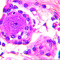
Thickening of the Schneiderian membrane (SM, mucosa of the maxillary sinus) appears in the paranasal sinus. Information on SM thickening is available for patients receiving sinus lift treatments, which is a risk factor for SM excretory dysfunction. However, more information is needed on the structure of the SM and the relationship between the maxilla sinus and palatine with the alveolar bone and the SM for dental implant treatment in the human maxilla. One hundred twenty-six sides of the maxilla from 71 cadavers were subjected to cone-beam CT (CBCT) analysis and macroscopic and immunohistochemical observations in this study. A thickened SM was mainly observed in the middle region of the basal layer of the maxillary sinus (MS). Strong calcitonin gene-related peptide (CGRP)-positive reactions were observed in the alveolar bone, oral mucosa, mucosa of the maxillary sinus, and trigeminal ganglion (TG) cells in dentulous samples compared with edentulous samples. TG cells play important roles in delivering CGRP through axons to the mucosal gland and in regulating the maxilla-related thickening of the SM. These data could help determine CGRP functions in the mucosal gland and bone formation between dentulous and edentulous samples and indicate that CGRP may pass from the TG to the maxillary sinus glands.

Dimensions

Altmetric

PlumX Metrics

Downloads

Download data is not yet available.

Citations

Beule AG. [Physiology and pathophysiology of respiratory mucosa of the nose and the paranasal sinuses].[Article in German]. Laryngorhinootologie 2010;89:S15-34.
Mygind N, Pedersen M, Nielsen MH. Morphology of the upper respiratory epithelium. DF Proctor and Andersen I, Editors. The Nose, upper airway physiology and the atmospheric environment. Amsterdam: Elsevier; 1982. p. 71-97.
Coleman H, Meer S, Altini M, Reyneke J, Becker P. Maxillary sinus pathology in 119 patients--a histopathologic study. SADJ 2005;140:142-5.
Manji A, Faucher J, Resnik RR, Suzuki JB. Prevalence of maxillary sinus pathology in patients considered for sinus augmentation procedures for dental implants. Implant Dent 2013;22:428-35. DOI: https://doi.org/10.1097/ID.0b013e31829d1a20
Ritter A, Rozendorn N, Avishai G, Rosenfeld E, Koren I, Soudry E. Preoperative maxillary sinus imaging and the outcome of sinus floor augmentation and dental implants in asymptomatic patients. Ann Otol Rhinol Laryngol 2020;129:209-15. DOI: https://doi.org/10.1177/0003489419883292
Ata-Ali J, Diago-Vilalta J-V, Melo M, Bagán L, Soldini M-C, Di-Nardo C, et al. What is the frequency of anatomical variations and pathological findings in maxillary sinuses among patients subjected to maxillofacial cone beam computed tomography? A systematic review. Med Oral Patol Oral Cir Bucal 2017;22:e400-9. DOI: https://doi.org/10.4317/medoral.21456
Munhoz L, Abdala Júnior R, Abdala R, Asaumi J, Arita ES. Diffusion-weighted magnetic resonance imaging in maxillary sinuses inflammatory diseases: Report of three cases and literature review. J Oral Maxillofac Res 2018;9:e4. DOI: https://doi.org/10.5037/jomr.2018.9204
Monje A, Monje-Gil F, Burgueño M, Gonzalez-Garcia R, Galindo-Moreno P, Wang HL. Incidence of and factors associated with sinus membrane perforation during maxillary sinus augmentation using the reamer drilling approach: A double-center case series. Int J Periodontics Restorative Dent 2016; 36:549-56. DOI: https://doi.org/10.11607/prd.2525
Insua A, Monje A, Chan HL, Zimmo N, Shaikh L, Wang HL. Accuracy of Schneiderian membrane thickness: a cone-beam computed tomography analysis with histological validation. Clin Oral Implants Res 2017; 28:654-661. DOI: https://doi.org/10.1111/clr.12856
Zimmo N, Insua A, Sinjab K, Chan HL, Shaikh L, Wang HL. Impact of sex, age, and season on sinus membrane thickness. Int J Oral Maxillofac Implants 2018;33:175-80. DOI: https://doi.org/10.11607/jomi.5924
Talo Yildirim T, Güncü GN, Colak M, Tözüm TF. The relationship between maxillary sinus lateral wall thickness, alveolar bone loss, and demographic variables: A cross-sectional cone-beam computerized tomography study. Med Princ Pract. 2019; 28:109-14. DOI: https://doi.org/10.1159/000494325
Bronzetti E, Artico M, Kovacs I, Felici LM, Magliulo G, Vignone D, et al. Expression of neurotransmitters and neurotrophins in neurogenic inflammation of the rat retina. Eur J Histochem 2007;51:251-60.
Sato I, Imura K, Miwa Y, Yoshida S, Sunohara M. Distributions of calcitonin gene-related peptide and substance P in the human maxillary sinus of Japanese cadavers. J Craniomaxillofac Surg 2012;40:e249-52. DOI: https://doi.org/10.1016/j.jcms.2011.10.027
Azuma Y, Sato I. The localization of calcitonin gene-related peptide in the human trigeminal ganglion and masseter muscle. Okajimas Folia Anat Jpn 2017;93:127-38. DOI: https://doi.org/10.2535/ofaj.93.127
Li J, Kreicbergs A, Bergström J, Stark A, Ahmed M. Site-specific CGRP innervation coincides with bone formation during fracture healing and modeling: A study in rat angulated tibia. J Orthop Res 2007;25:1204-12. DOI: https://doi.org/10.1002/jor.20406
Mitsuoka K, Kikutani T, Miwa Y, Sato I. Expression of CGRP neurotransmitter and vascular genesis marker mRNA is age-dependent in superior cervical ganglia of senescence-accelerated prone mice. Neurosci Lett 2018; 664:144-51. DOI: https://doi.org/10.1016/j.neulet.2017.11.034
Cassar-Malek I, Passelaigue F, Bernard C, Léger J, Hocquette JF. Target genes of myostatin loss-of-function in muscles of late bovine fetuses. BMC Genomics 2007;8:63. DOI: https://doi.org/10.1186/1471-2164-8-63
Ericson, S., Welander U. Local hyperplasia of the maxillary sinus mucosa after elimination of adjacent periapical osteitis. A follow-up study. Odontol Revy 1966;17:153-9.
Hauman CH, Chandler NP, Tong DC. Endodontic implications of the maxillary sinus: a review. Int Endod J 2002;35:127-41. DOI: https://doi.org/10.1046/j.0143-2885.2001.00524.x
Carmeli G, Artzi Z, Kozlovsky A, Segev Y, Landsberg R. Antral computerized tomography pre-operative evaluation: relationship between mucosal thickening and maxillary sinus function. Clin Oral Implants Res 2011;22:78-82. DOI: https://doi.org/10.1111/j.1600-0501.2010.01986.x
Sánchez-Pérez A, Boracchia AC, López-Jornet P, Boix-García P. Characterization of the maxillary sinus using cone beam computed tomography. A retrospective radiographic study. Implant Dent. 2016; 25:762-769. DOI: https://doi.org/10.1097/ID.0000000000000485
Panneton WM, Gan Q. Direct reticular projections of trigeminal sensory fibers immunoreactive to CGRP: potential monosynaptic somatoautonomic projections. Front Neurosci 2014;8:136.
Fang Z, Yang Q, Xiong W, Li GH, Liao H, Xiao J, et al. Effect of CGRP-adenoviral vector transduction on the osteoblastic differentiation of rat adipose-derived stem cells. PLoS One 2013;8:e72738. DOI: https://doi.org/10.1371/journal.pone.0072738
Wang J, Dong R, Zheng S. Roles of the inflammasome in the gut liver axis (Review). Mol Med Rep 2019;19:3-14. DOI: https://doi.org/10.1007/s00894-018-3886-2
Stjärne P, Lundblad L, Anggård A, Hökfelt T, Lundberg JM. Tachykinins and calcitonin gene-related peptide: co-existence in sensory nerves of the nasal mucosa and effects on blood flow. Cell Tissue Res 1989;256:439-46. DOI: https://doi.org/10.1007/BF00225591
Baraniuk JN, Lundgren JD, Goff J, Mullol J, Castellino S, Merida M, Shelhamer JH, Kaliner MA. Calcitonin gene-related peptide in human nasal mucosa. Am J Physiol 1990;258 L81-8. DOI: https://doi.org/10.1152/ajplung.1990.258.2.L81
Stjärne P, Lacroix JS, Anggård A, Lundberg JM. Compartment analysis of vascular effects of neuropeptides and capsaicin in the pig nasal mucosa. Acta Physiol Scand 1991;141:335-42. DOI: https://doi.org/10.1111/j.1748-1716.1991.tb09089.x
Lundberg JM. Peptidergic control of the autonomic regulation system in the orofacial region. Proc Fin Dent Soc 1989; 85:239-50.
Kuo HP, Rohde JA, Tokuyama K, Barnes PJ, Rogers DF. Capsaicin and sensory neuropeptide stimulation of goblet cell secretion in guinea-pig trachea. J Physiol 1990;431:629-41. DOI: https://doi.org/10.1113/jphysiol.1990.sp018351
Matran R. Neural control of lower airway vasculature. Involvement of classical transmitters and neuropeptides. Acta Phsiol 1991;601:1-54.
Irie K, Hara-Irie F, Ozawa H, Yajima T. Calcitonin gene-related peptide (CGRP)-containing nerve fibers in bone tissue and their involvement in bone remodeling. Microsc Res Tech 2002;58:85-90. DOI: https://doi.org/10.1002/jemt.10122
Nimigean V, Nimigean VR, Măru N, Sălăvăstru DI, Bădiţă D, Tuculină MJ. The maxillary sinus floor in the oral implantology. Rom J Morphol Embryol 2008;49:485-9.
Koppe T, Weigel C, Bärenklau M, Kaduk W, Bayerlein T, Gedrange T. Maxillary sinus pneumatization of an adult skull with an untreated bilateral cleft palate. J Craniomaxillofac Surg 2006;34:91-5. DOI: https://doi.org/10.1016/S1010-5182(06)60020-6
Heffner MA, Genetos DC, Christiansen BA. Bone adaptation to mechanical loading in a mouse model of reduced peripheral sensory nerve function. PLoS One 2017;12:e0187354. DOI: https://doi.org/10.1371/journal.pone.0187354
Damico JP, Ervolino E, Torres KR, Sabino Batagello D, Cruz-Rizzolo RJ, Aparecido Casatti C, Arruda Bauer J. Phenotypic alterations of neuropeptide Y and calcitonin gene-related peptide-containing neurons innervating the rat temporomandibular joint during carrageenan-induced arthritis. Eur J Histochem 2012;56:e31. DOI: https://doi.org/10.4081/ejh.2012.e31
Solar P, Geyerhofer U, Traxler H, Windisch A, Ulm C, Watzek G. Blood supply to the maxillary sinus relevant to sinus floor elevation procedures. Clin Oral Implants Res 1999;10:34-44. DOI: https://doi.org/10.1034/j.1600-0501.1999.100105.x
Miwa Y, Asaumi R, Kawai T, Maeda Y, Sato I. Morphological observation and CBCT of the bony canal structure of the groove and the location of blood vessels and nerves in the palatine of elderly human cadavers. Surg Radiol Anat 2018;40:199-206. DOI: https://doi.org/10.1007/s00276-017-1952-6
Kudo K, Tanaka K, Ambe K, Kawaai H, Yamazaki S. Immunohistochemical analysis of nerve distribution in mandible of rats. Anesth Prog 2019;66:87-93. DOI: https://doi.org/10.2344/anpr-66-01-10
Chan HL, Wang HL. Sinus pathology and anatomy in relation to complications in lateral window sinus augmentation. Implant Dent 2011;20:406-12. DOI: https://doi.org/10.1097/ID.0b013e3182341f79
Testori T, Tavelli L, Yu SH, Scaini R, Darnahal A, Wallace SS et al. Maxillary sinus elevation difficulty score with lateral wall technique. Int J Oral Maxillofac Implants 2020;35:631-8. DOI: https://doi.org/10.11607/jomi.8034

How to Cite

Matsuda, H., Sato, I., Asaumi, R., Omotehara, T., Kawata, S., Nagahori, K., Li, Z.-L., & Itoh, M. (2021). Comparison of CGRP distributions in the maxillary sinus and trigeminal ganglion between elderly dentulous and edentulous humans. European Journal of Histochemistry, 65(2). https://doi.org/10.4081/ejh.2021.3234