Quantitative, structural and molecular changes in neuroglia of aging mammals: A review

Accepted: 27 May 2021
HTML: 11
All claims expressed in this article are solely those of the authors and do not necessarily represent those of their affiliated organizations, or those of the publisher, the editors and the reviewers. Any product that may be evaluated in this article or claim that may be made by its manufacturer is not guaranteed or endorsed by the publisher.
Authors
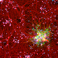
The neuroglia of the central and peripheral nervous systems undergo numerous changes during normal aging. Astrocytes become hypertrophic and accumulate intermediate filaments. Oligodendrocytes and Schwann cells undergo alterations that are often accompanied by degenerative changes to the myelin sheath. In microglia, proliferation in response to injury, motility of cell processes, ability to migrate to sites of neural injury, and phagocytic and autophagic capabilities are reduced. In sensory ganglia, the number and extent of gaps between perineuronal satellite cells – that leave the surfaces of sensory ganglion neurons directly exposed to basal lamina– increase significantly. The molecular profiles of neuroglia also change in old age, which, in view of the interactions between neurons and neuroglia, have negative consequences for important physiological processes in the nervous system. Since neuroglia actively participate in numerous nervous system processes, it is likely that not only neurons but also neuroglia will prove to be useful targets for interventions to prevent, reverse or slow the behavioral changes and cognitive decline that often accompany senescence.
How to Cite
PAGEPress has chosen to apply the Creative Commons Attribution NonCommercial 4.0 International License (CC BY-NC 4.0) to all manuscripts to be published.
Similar Articles
- Renata Tabola, Magdalena Zaremba-Czogalla, Dagmara Baczynska, Roberto Cirocchi, Kamila Stach, Krzysztof Grabowski, Katarzyna Augoff, Fibroblast activating protein-α expression in squamous cell carcinoma of the esophagus in primary and irradiated tumors: the use of archival FFPE material for molecular techniques , European Journal of Histochemistry: Vol. 61 No. 2 (2017)
- J. Rieger, P. Janczyk, H. Hünigen, J. Plendl, Enhancement of immunohistochemical detection of Salmonella in tissues of experimentally infected pigs , European Journal of Histochemistry: Vol. 59 No. 3 (2015)
- Bita Moudi, Zahra Heidari, Hamidreza Mahmoudzadeh-Sagheb, Seyed-Moayed Alavian, Kamran B. Lankarani, Parisa Farrokh, Jens Randel Nyengaard, Concomitant use of heat-shock protein 70, glutamine synthetase and glypican-3 is useful in diagnosis of HBV-related hepatocellular carcinoma with higher specificity and sensitivity , European Journal of Histochemistry: Vol. 62 No. 1 (2018)
- KH Cho, HS Lee, SK Ku, Changes in gastric endocrine cells in Balb/c mice bearing CT-26 carcinoma cells: an immunohistochemical study , European Journal of Histochemistry: Vol. 50 No. 4 (2006)
- H Ishida, M Kumakiri, K Ueda, LM Lao, M Yanagihara, K Asamoto, Y Imamura, S Noriki, M Fukuda, Comparative histochemical study of Bowen’s disease and actinic keratosis: preserved normal basal cells in Bowen’s disease , European Journal of Histochemistry: Vol. 45 No. 2 (2001)
- Z. Shen, Y. Ren, D. Ye, J. Guo, C. Kang, H. Ding, Significance and relationship between DJ-1 gene and survivin gene expression in laryngeal carcinoma , European Journal of Histochemistry: Vol. 55 No. 1 (2011)
- Giuliano Mazzini, The Feulgen reaction: from pink-magenta to rainbow fluorescent at the Maffo Vialli’s School of Histochemistry , European Journal of Histochemistry: Vol. 68 No. 1 (2024): 1954-2024: 70 Years of Histochemical Research
- I.C. Šoštarić-Zuckermann, K. Severin, M. Huzak, M. Hohšteter, A. Gudan Kurilj, B. Artuković, A. Džaja, Ž. Grabarević, Quantification of morphology of canine circumanal gland tumors: a fractal based study , European Journal of Histochemistry: Vol. 60 No. 2 (2016)
- S.A. Ferreira, J.L.A. Vasconcelos, R.C.W.C. Silva, C.L.B. Cavalcanti, C.L. Bezerra, M.J.B.M. Rêgo, E.I.C. Beltrão, Expression patterns of α2,3-Sialyltransferase I and α2,6-Sialyltransferase I in human cutaneous epithelial lesions , European Journal of Histochemistry: Vol. 57 No. 1 (2013)
- CarloAlberto Redi, Adult stem cells - Biology and methods of analysis , European Journal of Histochemistry: Vol. 55 No. 4 (2011)
<< < 35 36 37 38 39 40 41 42 43 44 > >>
You may also start an advanced similarity search for this article.

 https://doi.org/10.4081/ejh.2021.3249


