Quantitative, structural and molecular changes in neuroglia of aging mammals: A review

Accepted: 27 May 2021
HTML: 11
All claims expressed in this article are solely those of the authors and do not necessarily represent those of their affiliated organizations, or those of the publisher, the editors and the reviewers. Any product that may be evaluated in this article or claim that may be made by its manufacturer is not guaranteed or endorsed by the publisher.
Authors
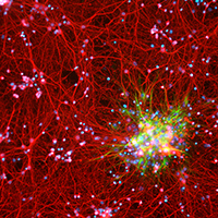
The neuroglia of the central and peripheral nervous systems undergo numerous changes during normal aging. Astrocytes become hypertrophic and accumulate intermediate filaments. Oligodendrocytes and Schwann cells undergo alterations that are often accompanied by degenerative changes to the myelin sheath. In microglia, proliferation in response to injury, motility of cell processes, ability to migrate to sites of neural injury, and phagocytic and autophagic capabilities are reduced. In sensory ganglia, the number and extent of gaps between perineuronal satellite cells – that leave the surfaces of sensory ganglion neurons directly exposed to basal lamina– increase significantly. The molecular profiles of neuroglia also change in old age, which, in view of the interactions between neurons and neuroglia, have negative consequences for important physiological processes in the nervous system. Since neuroglia actively participate in numerous nervous system processes, it is likely that not only neurons but also neuroglia will prove to be useful targets for interventions to prevent, reverse or slow the behavioral changes and cognitive decline that often accompany senescence.
How to Cite
PAGEPress has chosen to apply the Creative Commons Attribution NonCommercial 4.0 International License (CC BY-NC 4.0) to all manuscripts to be published.
Similar Articles
- Qinan Yin, Introduction to the Special Issue on Stem Cells and Regenerative Medicine , European Journal of Histochemistry: Vol. 64 No. s2 (2020): Special Collection on Stem Cells and Regenerative Medicine
- M Zhou, HJ He, R Suzuki, O Tanaka, M Sekiguchi, Y Yasuoka, K Kawahara, H Itoh, H Abe, Expression of ATP sensitive K+ channel subunit Kir6.1 in rat kidney , European Journal of Histochemistry: Vol. 51 No. 1 (2007)
- Francesca Scolari, Alessandro Girella, Anna Cleta Croce, Imaging and spectral analysis of autofluorescence patterns in larval head structures of mosquito vectors , European Journal of Histochemistry: Vol. 66 No. 4 (2022)
- Fengqin Yan, Hong Zhu, Yingxia He, Qinqin Wu, Xiaoyu Duan, Combination of tolvaptan and valsartan improves cardiac and renal functions in doxorubicin-induced heart failure in mice , European Journal of Histochemistry: Vol. 66 No. 4 (2022)
- C Pellicciari, MG Bottone, AI Scovassi, TE Martin, M Biggiogera, Rearrangement of nuclear ribonucleoproteins and extrusion of nucleolus-like bodies during apoptosis induced by hypertonic stress , European Journal of Histochemistry: Vol. 44 No. 3 (2000)
- ChenHui Zhu, LiJuan Lin, ChangQing Huang, ZhiHui Wu, Activation of Hedgehog pathway by circEEF2/miR-625-5p/TRPM2 axis promotes prostate cancer cell proliferation through mitochondrial stress , European Journal of Histochemistry: Vol. 68 No. 4 (2024)
- Manuela Monti, Microtubule dynamics , European Journal of Histochemistry: Vol. 56 No. 1 (2012)
- D Necchi, C Soldani, F Ronchetti, G Bernocchi, E Scherini, MPTP-induced increase in c-Fos- and c-Jun-like immunoreactivity in the monkey cerebellum , European Journal of Histochemistry: Vol. 48 No. 4 (2004)
- Shule Hou, Jiarui Chen, Jun Yang, Autophagy precedes apoptosis during degeneration of the Kölliker’s organ in the development of rat cochlea , European Journal of Histochemistry: Vol. 63 No. 2 (2019)
- A Zulli, LM Burrell, BF Buxton, DL Hare, ACE2 and AT4R are present in diseased human blood vessels , European Journal of Histochemistry: Vol. 52 No. 1 (2008)
<< < 68 69 70 71 72 73 74 75 76 77 > >>
You may also start an advanced similarity search for this article.

 https://doi.org/10.4081/ejh.2021.3249


