2D vs 3D morphological analysis of dorsal root ganglia in health and painful neuropathy

Accepted: 16 August 2021
Video 1: 177
Video 2: 193
HTML: 16
All claims expressed in this article are solely those of the authors and do not necessarily represent those of their affiliated organizations, or those of the publisher, the editors and the reviewers. Any product that may be evaluated in this article or claim that may be made by its manufacturer is not guaranteed or endorsed by the publisher.
Authors
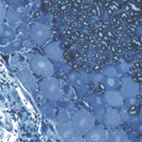
Dorsal root ganglia (DRGs) are clusters of sensory neurons that transmit the sensory information from the periphery to the central nervous system, and satellite glial cells (SGCs), their supporting trophic cells. Sensory neurons are pseudounipolar neurons with a heterogeneous neurochemistry reflecting their functional features. DRGs, not protected by the blood brain barrier, are vulnerable to stress and damage of different origin (i.e., toxic, mechanical, metabolic, genetic) that can involve sensory neurons, SGCs or, considering their intimate intercommunication, both cell populations. DRG damage, primary or secondary to nerve damage, produces a sensory peripheral neuropathy, characterized by neurophysiological abnormalities, numbness, paraesthesia and dysesthesia, tingling and burning sensations and neuropathic pain. DRG stress can be morphologically detected by light and electron microscope analysis with alterations in cell size (swelling/atrophy) and in different sub-cellular compartments (i.e., mitochondria, endoplasmic reticulum, and nucleus) of neurons and/or SGCs. In addition, neurochemical changes can be used to portray abnormalities of neurons and SGC. Conventional immunostaining, i.e., immunohistochemical detection of specific molecules in tissue slices can be employed to detect, localize and quantify particular markers of damage in neurons (i.e., nuclear expression ATF3) or SGCs (i.e., increased expression of GFAP), markers of apoptosis (i.e., caspases), markers of mitochondrial suffering and oxidative stress (i.e., 8-OHdG), markers of tissue inflammation (i.e., CD68 for macrophage infiltration), etc. However classical (2D) methods of immunostaining disrupt the overall organization of the DRG, thus resulting in the loss of some crucial information. Whole-mount (3D) methods have been recently developed to investigate DRG morphology and neurochemistry without tissue slicing, giving the opportunity to study the intimate relationship between SGCs and sensory neurons in health and disease. Here, we aim to compare classical (2D) vs whole-mount (3D) approaches to highlight “pros” and “cons” of the two methodologies when analysing neuropathy-induced alterations in DRGs.
How to Cite

This work is licensed under a Creative Commons Attribution-NonCommercial 4.0 International License.
PAGEPress has chosen to apply the Creative Commons Attribution NonCommercial 4.0 International License (CC BY-NC 4.0) to all manuscripts to be published.
Similar Articles
- Lan Wang, Zhenyu Fan, Haijin Wang, Shougui Xiang, Propofol alleviates M1 polarization and neuroinflammation of microglia in a subarachnoid hemorrhage model in vitro, by targeting the miR-140-5p/TREM-1/NF-κB signaling axis , European Journal of Histochemistry: Vol. 68 No. 3 (2024)
- Elva I. Cortés Gutiérrez, Catalina García-Vielma, Adriana Aguilar-Lemarroy, Veronica Vallejo-Ruíz, Patricia Piña-Sánchez, Pablo Zapata-Benavides, Jaime Gosalvez, Expression of the HPV18/E6 oncoprotein induces DNA damage , European Journal of Histochemistry: Vol. 61 No. 2 (2017)
- Shiyuan Chen, Longfei Zhang, Benchi Feng, Wei Wang, Delang Liu, Xinyu Zhao, Chaowen Yu, Xiaogao Wang, Yong Gao, MiR-550a-3p restores damaged vascular smooth muscle cells by inhibiting thrombomodulin in an in vitro atherosclerosis model , European Journal of Histochemistry: Vol. 66 No. 3 (2022)
- R. M. Ruggeri, E. Vitarelli, G. Barresi, F. Trimarchi, S. Benvenga, M. Trovato, The tyrosine kinase receptor c-met, its cognate ligand HGF and the tyrosine kinase receptor trasducers STAT3, PI3K and RHO in thyroid nodules associated with Hashimoto's thyroiditis: an immunohistochemical characterization , European Journal of Histochemistry: Vol. 54 No. 2 (2010)
- M. Kawashima, K. Imura, I. Sato, Topographical organization of TRPV1-immunoreactive epithelium and CGRP-immunoreactive nerve terminals in rodent tongue , European Journal of Histochemistry: Vol. 56 No. 2 (2012)
- Yangying Peng, Shaojie Ding, Ping Xu, Xueyan Zhang, Jianzhang Wang, Tiantian Li, Liyun Liao, Xinmei Zhang, CCL18 promotes endometriosis by increasing endometrial cell migration and neuroangiogenesis , European Journal of Histochemistry: Vol. 68 No. 3 (2024)
- A. Porzionato, M. M. Sfriso, V. Macchi, A. Rambaldo, G. Lago, L. Lancerotto, V. Vindigni, R. De Caro, Decellularized omentum as novel biologic scaffold for reconstructive surgery and regenerative medicine , European Journal of Histochemistry: Vol. 57 No. 1 (2013)
- Marina Boido, Elena De Amicis, Katia Mareschi, Franca Fagioli, Alessandro Vercelli, Organotypic spinal cord cultures: An in vitro 3D model to preliminary screen treatments for spinal muscular atrophy , European Journal of Histochemistry: Vol. 65 No. s1 (2021): Special Collection on Advances in Neuromorphology in Health and Disease
- Andreea Cioca, Amalia R. Ceausu, Irina Marin, Marius Raica, Anca M. Cimpean, The multifaceted role of podoplanin expression in hepatocellular carcinoma , European Journal of Histochemistry: Vol. 61 No. 1 (2017)
- Liuchang Feng, Zaoqiang Lin, Zeyong Tang, Lin Zhu, Shu Xu, Xi Tan, Xinyuan Wang, Jianling Mai, Qinxiang Tan, Emodin improves renal fibrosis in chronic kidney disease by regulating mitochondrial homeostasis through the mediation of peroxisome proliferator-activated receptor-gamma coactivator-1 alpha (PGC-1α) , European Journal of Histochemistry: Vol. 68 No. 2 (2024)
<< < 11 12 13 14 15 16 17 18 19 20 > >>
You may also start an advanced similarity search for this article.

 https://doi.org/10.4081/ejh.2021.3276


