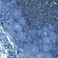
2D vs 3D morphological analysis of dorsal root ganglia in health and painful neuropathy

Accepted: 16 August 2021
Video 1: 177
Video 2: 193
HTML: 16
All claims expressed in this article are solely those of the authors and do not necessarily represent those of their affiliated organizations, or those of the publisher, the editors and the reviewers. Any product that may be evaluated in this article or claim that may be made by its manufacturer is not guaranteed or endorsed by the publisher.
Authors
Dorsal root ganglia (DRGs) are clusters of sensory neurons that transmit the sensory information from the periphery to the central nervous system, and satellite glial cells (SGCs), their supporting trophic cells. Sensory neurons are pseudounipolar neurons with a heterogeneous neurochemistry reflecting their functional features. DRGs, not protected by the blood brain barrier, are vulnerable to stress and damage of different origin (i.e., toxic, mechanical, metabolic, genetic) that can involve sensory neurons, SGCs or, considering their intimate intercommunication, both cell populations. DRG damage, primary or secondary to nerve damage, produces a sensory peripheral neuropathy, characterized by neurophysiological abnormalities, numbness, paraesthesia and dysesthesia, tingling and burning sensations and neuropathic pain. DRG stress can be morphologically detected by light and electron microscope analysis with alterations in cell size (swelling/atrophy) and in different sub-cellular compartments (i.e., mitochondria, endoplasmic reticulum, and nucleus) of neurons and/or SGCs. In addition, neurochemical changes can be used to portray abnormalities of neurons and SGC. Conventional immunostaining, i.e., immunohistochemical detection of specific molecules in tissue slices can be employed to detect, localize and quantify particular markers of damage in neurons (i.e., nuclear expression ATF3) or SGCs (i.e., increased expression of GFAP), markers of apoptosis (i.e., caspases), markers of mitochondrial suffering and oxidative stress (i.e., 8-OHdG), markers of tissue inflammation (i.e., CD68 for macrophage infiltration), etc. However classical (2D) methods of immunostaining disrupt the overall organization of the DRG, thus resulting in the loss of some crucial information. Whole-mount (3D) methods have been recently developed to investigate DRG morphology and neurochemistry without tissue slicing, giving the opportunity to study the intimate relationship between SGCs and sensory neurons in health and disease. Here, we aim to compare classical (2D) vs whole-mount (3D) approaches to highlight “pros” and “cons” of the two methodologies when analysing neuropathy-induced alterations in DRGs.
How to Cite

This work is licensed under a Creative Commons Attribution-NonCommercial 4.0 International License.
PAGEPress has chosen to apply the Creative Commons Attribution NonCommercial 4.0 International License (CC BY-NC 4.0) to all manuscripts to be published.
Similar Articles
- E. Carabajal, N. Massari, M. Croci, D. J. Martinel Lamas, J. P. Prestifilippo, R. M. Bergoc, E. S. Rivera, V. A. Medina, Radioprotective potential of histamine on rat small intestine and uterus , European Journal of Histochemistry: Vol. 56 No. 4 (2012)
- C. Severi, R. Sferra, A. Scirocco, A. Vetuschi, N. Pallotta, A. Pronio, R. Caronna, G. Di Rocco, E. Gaudio, E. Corazziari, P. Onori, Contribution of intestinal smooth muscle to Crohn's disease fibrogenesis , European Journal of Histochemistry: Vol. 58 No. 4 (2014)
- G. Musumeci, C. Loreto, M.L. Carnazza, F. Coppolino, V. Cardile, R. Leonardi, Lubricin is expressed in chondrocytes derived from osteoarthritic cartilage encapsulated in poly(ethylene glycol) diacrylate scaffold , European Journal of Histochemistry: Vol. 55 No. 3 (2011)
- Q. Li, J. Weng, H. Zhang, L. Lu, X. Ma, Q. Wang, H. Cao, S. Liu, M. Xu, Q. Weng, G. Watanabe, K. Taya, Immunohistochemical evidence: testicular and scented glandular androgen synthesis in muskrats (Ondatra zibethicus) during the breeding season , European Journal of Histochemistry: Vol. 55 No. 4 (2011)
- Francesca Diomede, Maria Zingariello, Marcos F.X.B. Cavalcanti, Ilaria Merciaro, Jacopo Pizzicannella, Natalia de Isla, Sergio Caputi, Patrizia Ballerini, Oriana Trubiani, MyD88/ERK/NFkB pathways and pro-inflammatory cytokines release in periodontal ligament stem cells stimulated by Porphyromonas gingivalis , European Journal of Histochemistry: Vol. 61 No. 2 (2017)
- Zhijiang Chen, Huili Wang, Bin Hu, Xinxin Chen, Meiyu Zheng, Lili Liang, Juanjuan Lyu, Qiyi Zeng, Transcription factor nuclear factor erythroid 2 p45-related factor 2 (NRF2) ameliorates sepsis-associated acute kidney injury by maintaining mitochondrial homeostasis and improving the mitochondrial function , European Journal of Histochemistry: Vol. 66 No. 3 (2022)
- Letizia Ferroni, Chiara Gardin, Andrea De Pieri, Maria Sambataro, Elena Seganfreddo, Chiara Goretti, Elisabetta Iacopi, Barbara Zavan, Alberto Piaggesi, Treatment of diabetic foot ulcers with Therapeutic Magnetic Resonance (TMR®) improves the quality of granulation tissue , European Journal of Histochemistry: Vol. 61 No. 3 (2017)
- Y. Zhu, D. Ning, F. Wang, C. Liu, Y. Xu, X. Jia, D. Zhu, Effect of thyroxine on munc-18 and syntaxin-1 expression in dorsal hippocampus of adult-onset hypothyroid rats , European Journal of Histochemistry: Vol. 56 No. 2 (2012)
- E. Akat, H. Arıkan, B. Göçmen, Histochemical and biometric study of the gastrointestinal system of Hyla orientalis (Bedriaga, 1890) (Anura, Hylidae) , European Journal of Histochemistry: Vol. 58 No. 4 (2014)
- Peipei Lu, Shuxiang Li, Caoyang Zhang, Xinyi Jiang, Jinghua Xiang, Hong Xu, Jian Dong, Kun Wang, Yuhua Shi, Spinosin ameliorates osteoarthritis through enhancing the Nrf2/HO-1 signaling pathway , European Journal of Histochemistry: Vol. 68 No. 2 (2024)
<< < 13 14 15 16 17 18 19 20 21 22 > >>
You may also start an advanced similarity search for this article.

 https://doi.org/10.4081/ejh.2021.3276


