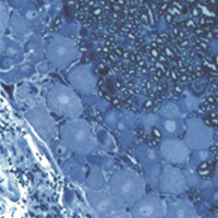
2D vs 3D morphological analysis of dorsal root ganglia in health and painful neuropathy

Accepted: 16 August 2021
Video 1: 177
Video 2: 193
HTML: 16
All claims expressed in this article are solely those of the authors and do not necessarily represent those of their affiliated organizations, or those of the publisher, the editors and the reviewers. Any product that may be evaluated in this article or claim that may be made by its manufacturer is not guaranteed or endorsed by the publisher.
Authors
Dorsal root ganglia (DRGs) are clusters of sensory neurons that transmit the sensory information from the periphery to the central nervous system, and satellite glial cells (SGCs), their supporting trophic cells. Sensory neurons are pseudounipolar neurons with a heterogeneous neurochemistry reflecting their functional features. DRGs, not protected by the blood brain barrier, are vulnerable to stress and damage of different origin (i.e., toxic, mechanical, metabolic, genetic) that can involve sensory neurons, SGCs or, considering their intimate intercommunication, both cell populations. DRG damage, primary or secondary to nerve damage, produces a sensory peripheral neuropathy, characterized by neurophysiological abnormalities, numbness, paraesthesia and dysesthesia, tingling and burning sensations and neuropathic pain. DRG stress can be morphologically detected by light and electron microscope analysis with alterations in cell size (swelling/atrophy) and in different sub-cellular compartments (i.e., mitochondria, endoplasmic reticulum, and nucleus) of neurons and/or SGCs. In addition, neurochemical changes can be used to portray abnormalities of neurons and SGC. Conventional immunostaining, i.e., immunohistochemical detection of specific molecules in tissue slices can be employed to detect, localize and quantify particular markers of damage in neurons (i.e., nuclear expression ATF3) or SGCs (i.e., increased expression of GFAP), markers of apoptosis (i.e., caspases), markers of mitochondrial suffering and oxidative stress (i.e., 8-OHdG), markers of tissue inflammation (i.e., CD68 for macrophage infiltration), etc. However classical (2D) methods of immunostaining disrupt the overall organization of the DRG, thus resulting in the loss of some crucial information. Whole-mount (3D) methods have been recently developed to investigate DRG morphology and neurochemistry without tissue slicing, giving the opportunity to study the intimate relationship between SGCs and sensory neurons in health and disease. Here, we aim to compare classical (2D) vs whole-mount (3D) approaches to highlight “pros” and “cons” of the two methodologies when analysing neuropathy-induced alterations in DRGs.
How to Cite

This work is licensed under a Creative Commons Attribution-NonCommercial 4.0 International License.
PAGEPress has chosen to apply the Creative Commons Attribution NonCommercial 4.0 International License (CC BY-NC 4.0) to all manuscripts to be published.
Similar Articles
- Hao Li, Junliang Chen, Wenjun You, Yizhen Xu, Yaqiong Ye, Haiquan Zhao, Junxing Li, Hui Zhang, Developmental characteristics of cutaneous telocytes in late embryos of the silky fowl , European Journal of Histochemistry: Vol. 68 No. 4 (2024)
- N. Kiga, I. Tojyo, T. Matsumoto, Y. Hiraishi, Y. Shinohara, S. Fujita, Expression of lumican related to CD34 and VEGF in the articular disc of the human temporomandibular joint. , European Journal of Histochemistry: Vol. 54 No. 3 (2010)
- P. Demurtas, M. Corrias, I. Zucca, C. Maxia, F. Piras, P. Sirigu, M.T. Perra, Angiotensin II: immunohistochemical study in Sardinian pterygium , European Journal of Histochemistry: Vol. 58 No. 3 (2014)
- W. Meyer, J. Kacza, I. N. Hornickel, B. Schoennagel, Immunolocalization of succinate dehydrogenase in the esophagus epithelium of domesticated mammals , European Journal of Histochemistry: Vol. 57 No. 2 (2013)
- Karel Smetana, Dana Mikulenková, Josef Karban, Marek Trněný, To the ring-shaped nucleolus seen by microscopy using human lymphocytes of blood donors and chronic lymphocytic leukemia patients , European Journal of Histochemistry: Vol. 68 No. 3 (2024)
- S. Nemolato, J. Ekstrom, T. Cabras, C. Gerosa, D. Fanni, E. Di Felice, A. Locci, I. Messana, M. Castagnola, G. Faa, Immunoreactivity for thymosin beta 4 and thymosin beta 10 in the adult rat oro-gastro-intestinal tract , European Journal of Histochemistry: Vol. 57 No. 2 (2013)
- Yan Long, Yan Zhao, Xiaoqing Ma, Ya Zeng, Tian Hu, Weijie Wu, Chongtian Deng, Jinyue Hu, Yueming Shen, Endoplasmic reticulum stress contributed to inflammatory bowel disease by activating p38 MAPK pathway , European Journal of Histochemistry: Vol. 66 No. 2 (2022)
- M. Lehmann, F. Martin, K. Mannigel, K. Kaltschmidt, U. Sack, U. Anderer, Three-dimensional scaffold-free fusion culture: the way to enhance chondrogenesis of in vitro propagated human articular chondrocytes , European Journal of Histochemistry: Vol. 57 No. 4 (2013)
- T. Karaca, Y. Hulya Uz, R. Karabacak, I. Karaboga, S. Demirtas, A. Cagatay Cicek, Effects of hyperthyroidism on expression of vascular endothelial growth factor (VEGF) and apoptosis in fetal adrenal glands , European Journal of Histochemistry: Vol. 59 No. 4 (2015)
- Yuuki Maeda, Yoko Miwa, Iwao Sato, Expression of CGRP, vasculogenesis and osteogenesis associated mRNAs in the developing mouse mandible and tibia , European Journal of Histochemistry: Vol. 61 No. 1 (2017)
<< < 21 22 23 24 25 26 27 28 29 30 > >>
You may also start an advanced similarity search for this article.

 https://doi.org/10.4081/ejh.2021.3276


