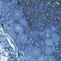
2D vs 3D morphological analysis of dorsal root ganglia in health and painful neuropathy

Accepted: 16 August 2021
Video 1: 177
Video 2: 193
HTML: 16
All claims expressed in this article are solely those of the authors and do not necessarily represent those of their affiliated organizations, or those of the publisher, the editors and the reviewers. Any product that may be evaluated in this article or claim that may be made by its manufacturer is not guaranteed or endorsed by the publisher.
Authors
Dorsal root ganglia (DRGs) are clusters of sensory neurons that transmit the sensory information from the periphery to the central nervous system, and satellite glial cells (SGCs), their supporting trophic cells. Sensory neurons are pseudounipolar neurons with a heterogeneous neurochemistry reflecting their functional features. DRGs, not protected by the blood brain barrier, are vulnerable to stress and damage of different origin (i.e., toxic, mechanical, metabolic, genetic) that can involve sensory neurons, SGCs or, considering their intimate intercommunication, both cell populations. DRG damage, primary or secondary to nerve damage, produces a sensory peripheral neuropathy, characterized by neurophysiological abnormalities, numbness, paraesthesia and dysesthesia, tingling and burning sensations and neuropathic pain. DRG stress can be morphologically detected by light and electron microscope analysis with alterations in cell size (swelling/atrophy) and in different sub-cellular compartments (i.e., mitochondria, endoplasmic reticulum, and nucleus) of neurons and/or SGCs. In addition, neurochemical changes can be used to portray abnormalities of neurons and SGC. Conventional immunostaining, i.e., immunohistochemical detection of specific molecules in tissue slices can be employed to detect, localize and quantify particular markers of damage in neurons (i.e., nuclear expression ATF3) or SGCs (i.e., increased expression of GFAP), markers of apoptosis (i.e., caspases), markers of mitochondrial suffering and oxidative stress (i.e., 8-OHdG), markers of tissue inflammation (i.e., CD68 for macrophage infiltration), etc. However classical (2D) methods of immunostaining disrupt the overall organization of the DRG, thus resulting in the loss of some crucial information. Whole-mount (3D) methods have been recently developed to investigate DRG morphology and neurochemistry without tissue slicing, giving the opportunity to study the intimate relationship between SGCs and sensory neurons in health and disease. Here, we aim to compare classical (2D) vs whole-mount (3D) approaches to highlight “pros” and “cons” of the two methodologies when analysing neuropathy-induced alterations in DRGs.
How to Cite

This work is licensed under a Creative Commons Attribution-NonCommercial 4.0 International License.
PAGEPress has chosen to apply the Creative Commons Attribution NonCommercial 4.0 International License (CC BY-NC 4.0) to all manuscripts to be published.
Similar Articles
- M. Chiusa, F. Timolati, J.C. Perriard, T.M. Suter, C. Zuppinger, Sodium nitroprusside induces cell death and cytoskeleton degradation in adult rat cardiomyocytes in vitro: implications for anthracycline-induced cardiotoxicity , European Journal of Histochemistry: Vol. 56 No. 2 (2012)
- M. Salemi, A. E. Calogero, G. Zaccarello, R. Castiglione, A. Cosentino, C. Campagna, E. Vicari, G. Rappazzo, Expression of SPANX proteins in normal prostatic tissue and in prostate cancer , European Journal of Histochemistry: Vol. 54 No. 3 (2010)
- Cecilia Dall'Aglio, Angela Polisca, Maria Grazia Cappai, Francesca Mercati, Alessandro Troisi, Carolina Pirino, Paola Scocco, Margherita Maranesi, Immunohistochemistry detected and localized cannabinoid receptor type 2 in bovine fetal pancreas at late gestation , European Journal of Histochemistry: Vol. 61 No. 1 (2017)
- Y. Liu, J. Weng, S. Huang, Y. Shen, X. Sheng, Y. Han, M. Xu, Q. Weng, Immunoreactivities of PPARγ2, leptin and leptin receptor in oviduct of Chinese brown frog during breeding period and pre-hibernation , European Journal of Histochemistry: Vol. 58 No. 3 (2014)
- Andrea Amaroli, Sara Ferrando, Marina Pozzolini, Lorenzo Gallus, Steven Parker, Stefano Benedicenti, The earthworm Dendrobaena veneta (Annelida): A new experimental-organism for photobiomodulation and wound healing , European Journal of Histochemistry: Vol. 62 No. 1 (2018)
- A. Vetuschi, A. D'Alfonso, R. Sferra, D. Zanelli, S. Pompili, F. Patacchiola, E. Gaudio, G. Carta, Changes in muscularis propria of anterior vaginal wall in women with pelvic organ prolapse , European Journal of Histochemistry: Vol. 60 No. 1 (2016)
- Lucia Aidos, Luisa M. Pinheiro Valente, Vera Sousa, Marco Lanfranchi, Cinzia Domeneghini, Alessia Di Giancamillo, Effects of different rearing temperatures on muscle development and stress response in the early larval stages of Acipenser baerii , European Journal of Histochemistry: Vol. 61 No. 4 (2017)
- Filippo Vernia, Tiziana Tatti, Stefano Necozione, Annalisa Capannolo, Nicola Cesaro, Marco Magistroni, Marco Valvano, Simona Pompili, Roberta Sferra, Antonella Vetuschi, Giovanni Latella, Is mastocytic colitis a specific clinical-pathological entity? , European Journal of Histochemistry: Vol. 66 No. 4 (2022)
- Qingjing Gao, Wenqian Xie, Wenjing Lu, Yuning Liu, Haolin Zhang, Yingying Han, Qiang Weng, Seasonal patterns of prolactin, prolactin receptor, and STAT5 expression in the ovaries of wild ground squirrels (Citellus dauricus Brandt) , European Journal of Histochemistry: Vol. 67 No. 4 (2023)
- E. Lacunza, V. Ferretti, C. Barbeito, A. Segal-Eiras, M. V. Croce, Immunohistochemical evidence of Muc1 expression during rat embryonic development , European Journal of Histochemistry: Vol. 54 No. 4 (2010)
<< < 25 26 27 28 29 30 31 32 33 34 > >>
You may also start an advanced similarity search for this article.

 https://doi.org/10.4081/ejh.2021.3276


