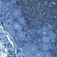
2D vs 3D morphological analysis of dorsal root ganglia in health and painful neuropathy

Accepted: 16 August 2021
Video 1: 177
Video 2: 193
HTML: 16
All claims expressed in this article are solely those of the authors and do not necessarily represent those of their affiliated organizations, or those of the publisher, the editors and the reviewers. Any product that may be evaluated in this article or claim that may be made by its manufacturer is not guaranteed or endorsed by the publisher.
Authors
Dorsal root ganglia (DRGs) are clusters of sensory neurons that transmit the sensory information from the periphery to the central nervous system, and satellite glial cells (SGCs), their supporting trophic cells. Sensory neurons are pseudounipolar neurons with a heterogeneous neurochemistry reflecting their functional features. DRGs, not protected by the blood brain barrier, are vulnerable to stress and damage of different origin (i.e., toxic, mechanical, metabolic, genetic) that can involve sensory neurons, SGCs or, considering their intimate intercommunication, both cell populations. DRG damage, primary or secondary to nerve damage, produces a sensory peripheral neuropathy, characterized by neurophysiological abnormalities, numbness, paraesthesia and dysesthesia, tingling and burning sensations and neuropathic pain. DRG stress can be morphologically detected by light and electron microscope analysis with alterations in cell size (swelling/atrophy) and in different sub-cellular compartments (i.e., mitochondria, endoplasmic reticulum, and nucleus) of neurons and/or SGCs. In addition, neurochemical changes can be used to portray abnormalities of neurons and SGC. Conventional immunostaining, i.e., immunohistochemical detection of specific molecules in tissue slices can be employed to detect, localize and quantify particular markers of damage in neurons (i.e., nuclear expression ATF3) or SGCs (i.e., increased expression of GFAP), markers of apoptosis (i.e., caspases), markers of mitochondrial suffering and oxidative stress (i.e., 8-OHdG), markers of tissue inflammation (i.e., CD68 for macrophage infiltration), etc. However classical (2D) methods of immunostaining disrupt the overall organization of the DRG, thus resulting in the loss of some crucial information. Whole-mount (3D) methods have been recently developed to investigate DRG morphology and neurochemistry without tissue slicing, giving the opportunity to study the intimate relationship between SGCs and sensory neurons in health and disease. Here, we aim to compare classical (2D) vs whole-mount (3D) approaches to highlight “pros” and “cons” of the two methodologies when analysing neuropathy-induced alterations in DRGs.
How to Cite

This work is licensed under a Creative Commons Attribution-NonCommercial 4.0 International License.
PAGEPress has chosen to apply the Creative Commons Attribution NonCommercial 4.0 International License (CC BY-NC 4.0) to all manuscripts to be published.
Similar Articles
- Cheng Chen, Yan Huang, Pingping Xia, Fan Zhang, Longyan Li, E Wang, Qulian Guo, Zhi Ye, Long noncoding RNA Meg3 mediates ferroptosis induced by oxygen and glucose deprivation combined with hyperglycemia in rat brain microvascular endothelial cells, through modulating the p53/GPX4 axis , European Journal of Histochemistry: Vol. 65 No. 3 (2021)
- Xiaoying Yang, Xuhao Liu, Fengcheng Song, Hao Wei, Fuli Gao, Haolin Zhang, Yingying Han, Qiang Weng, Zhengrong Yuan, Seasonal expressions of GPR41 and GPR43 in the colon of the wild ground squirrels (Spermophilus dauricus) , European Journal of Histochemistry: Vol. 66 No. 1 (2022)
- M. Malatesta, C. Zancanaro, M. Costanzo, B. Cisterna, C. Pellicciari, Simultaneous ultrastructural analysis of fluorochrome-photoconverted diaminobenzidine and gold immunolabeling in cultured cells , European Journal of Histochemistry: Vol. 57 No. 3 (2013)
- You-Jie Liu, Hua-Jun Wang, Zhao-Wen Xue, Lek-Hang Cheang, Man-Seng Tam, Ri-Wang Li, Jie-Ruo Li, Hui-Ge Hou, Xiao-Fei Zheng, Long noncoding RNA H19 accelerates tenogenic differentiation by modulating miR-140-5p/VEGFA signaling , European Journal of Histochemistry: Vol. 65 No. 3 (2021)
- S. Grecchi, M. Malatesta, Visualizing endocytotic pathways at transmission electron microscopy via diaminobenzidine photo-oxidation by a fluorescent cell-membrane dye , European Journal of Histochemistry: Vol. 58 No. 4 (2014)
- G. Laguna Hernández, A.E. Brechú-Franco, I. De la Cruz-Chacón, A.R. González-Esquinca, Histochemical detection of acetogenins and storage molecules in the endosperm of Annona macroprophyllata Donn Sm. seeds , European Journal of Histochemistry: Vol. 59 No. 3 (2015)
- F. Mercati, L. Pascucci, P. Ceccarelli, C. Dall’Aglio, V. Pedini, A.M. Gargiulo, Expression of mesenchymal stem cell marker CD90 on dermal sheath cells of the anagen hair follicle in canine species , European Journal of Histochemistry: Vol. 53 No. 3 (2009)
- V. Poletto, V. Galimberti, G. Guerra, V. Rosti, F. Moccia, M. Biggiogera, Fine structural detection of calcium ions by photoconversion , European Journal of Histochemistry: Vol. 60 No. 3 (2016)
- A.K. Lindström, D. Hellberg, Immunohistochemical LRIG3 expression in cervical intraepithelial neoplasia and invasive squamous cell cervical cancer: association with expression of tumor markers, hormones, high-risk HPV-infection, smoking and patient outcome , European Journal of Histochemistry: Vol. 58 No. 2 (2014)
- Haixia Li, Ning Sun, Yaqiao Zhu, Wei Wang, Meihong Cai, Xiaohuan Luo, Wei Xia, Song Quan, Growth hormone inhibits the JAK/STAT3 pathway by regulating SOCS1 in endometrial cells in vitro: a clue to enhance endometrial receptivity in recurrent implantation failure , European Journal of Histochemistry: Vol. 67 No. 1 (2023)
<< < 27 28 29 30 31 32 33 34 35 36 > >>
You may also start an advanced similarity search for this article.

 https://doi.org/10.4081/ejh.2021.3276


