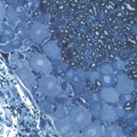
2D vs 3D morphological analysis of dorsal root ganglia in health and painful neuropathy

Accepted: 16 August 2021
Video 1: 177
Video 2: 193
HTML: 16
All claims expressed in this article are solely those of the authors and do not necessarily represent those of their affiliated organizations, or those of the publisher, the editors and the reviewers. Any product that may be evaluated in this article or claim that may be made by its manufacturer is not guaranteed or endorsed by the publisher.
Authors
Dorsal root ganglia (DRGs) are clusters of sensory neurons that transmit the sensory information from the periphery to the central nervous system, and satellite glial cells (SGCs), their supporting trophic cells. Sensory neurons are pseudounipolar neurons with a heterogeneous neurochemistry reflecting their functional features. DRGs, not protected by the blood brain barrier, are vulnerable to stress and damage of different origin (i.e., toxic, mechanical, metabolic, genetic) that can involve sensory neurons, SGCs or, considering their intimate intercommunication, both cell populations. DRG damage, primary or secondary to nerve damage, produces a sensory peripheral neuropathy, characterized by neurophysiological abnormalities, numbness, paraesthesia and dysesthesia, tingling and burning sensations and neuropathic pain. DRG stress can be morphologically detected by light and electron microscope analysis with alterations in cell size (swelling/atrophy) and in different sub-cellular compartments (i.e., mitochondria, endoplasmic reticulum, and nucleus) of neurons and/or SGCs. In addition, neurochemical changes can be used to portray abnormalities of neurons and SGC. Conventional immunostaining, i.e., immunohistochemical detection of specific molecules in tissue slices can be employed to detect, localize and quantify particular markers of damage in neurons (i.e., nuclear expression ATF3) or SGCs (i.e., increased expression of GFAP), markers of apoptosis (i.e., caspases), markers of mitochondrial suffering and oxidative stress (i.e., 8-OHdG), markers of tissue inflammation (i.e., CD68 for macrophage infiltration), etc. However classical (2D) methods of immunostaining disrupt the overall organization of the DRG, thus resulting in the loss of some crucial information. Whole-mount (3D) methods have been recently developed to investigate DRG morphology and neurochemistry without tissue slicing, giving the opportunity to study the intimate relationship between SGCs and sensory neurons in health and disease. Here, we aim to compare classical (2D) vs whole-mount (3D) approaches to highlight “pros” and “cons” of the two methodologies when analysing neuropathy-induced alterations in DRGs.
How to Cite

This work is licensed under a Creative Commons Attribution-NonCommercial 4.0 International License.
PAGEPress has chosen to apply the Creative Commons Attribution NonCommercial 4.0 International License (CC BY-NC 4.0) to all manuscripts to be published.
Similar Articles
- H. Huang, W. Wang, P. Liu, Y. Jiang, Y. Zhao, H. Wei, W. Niu, TRPC1 expression and distribution in rat hearts , European Journal of Histochemistry: Vol. 53 No. 4 (2009)
- S Passinen, T Ylikomi, Evidence for the existence of an oligomeric, non-DNA-binding complex of the progesterone receptor in the cytoplasm , European Journal of Histochemistry: Vol. 47 No. 3 (2003)
- A Alunni, S Vaccari, S Torcia, Characterization of glial fibrillary acidic protein and astroglial architecture in the brain of a continuously growing fish, the rainbow trout , European Journal of Histochemistry: Vol. 49 No. 2 (2005)
- T. Petr, V. Å mÃd, J. Å mÃdová, H. Hůlková, M. Jirkovská, M. Elleder, L. Muchová, L. Vitek, F. Å mÃd, Histochemical detection of GM1 ganglioside using cholera toxin-B subunit. Evaluation of critical factors optimal for in situ detection with special emphasis to acetone pre-extraction , European Journal of Histochemistry: Vol. 54 No. 2 (2010)
- G. Radaelli, C. Poltronieri, C. Simontacchi, E. Negrato, F. Pascoli, A. Libertini, D. Bertotto, Immunohistochemical localization of IGF-I, IGF-II and MSTN proteins during development of triploid sea bass (Dicentrarchus labrax) , European Journal of Histochemistry: Vol. 54 No. 2 (2010)
- Yao Le, Zhijun Wang, Qian Zhang, Ling Miao, Xiaohong Wang, Guorong Han, Study on the mechanism of Shenling Baizhu powder on the pathogenesis of pregnancy complicated with non-alcoholic fatty liver, based on PI3K/AKT/mTOR signal pathway , European Journal of Histochemistry: Vol. 68 No. 3 (2024)
- Lalith Prabha Ethiraj, En Lei Samuel Fong, Ranran Liu, Madelynn Chan, Christoph Winkler, Tom James Carney, Colorimetric and fluorescent TRAP assays for visualising and quantifying fish osteoclast activity , European Journal of Histochemistry: Vol. 66 No. 2 (2022)
- S Burattini, P Ferri, M Battistelli, R Curci, F Luchetti, E Falcieri, C2C12 murine myoblasts as a model of skeletal muscle development: morpho-functional characterization , European Journal of Histochemistry: Vol. 48 No. 3 (2004)
- Mihály Kálmán, Erzsébet Oszwald, István Adorján, Appearance of β-dystroglycan precedes the formation of glio-vascular end-feet in developing rat brain , European Journal of Histochemistry: Vol. 62 No. 2 (2018)
- Elena Pompili, Viviana Ciraci, Stefano Leone, Valerio De Franchis, Pietro Familiari, Roberto Matassa, Giuseppe Familiari, Ada Maria Tata, Lorenzo Fumagalli, Cinzia Fabrizi, Thrombin regulates the ability of Schwann cells to support neuritogenesis and to maintain the integrity of the nodes of Ranvier , European Journal of Histochemistry: Vol. 64 No. 2 (2020)
<< < 49 50 51 52 53 54 55 56 57 58 > >>
You may also start an advanced similarity search for this article.

 https://doi.org/10.4081/ejh.2021.3276


