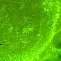
Organotypic spinal cord cultures: An in vitro 3D model to preliminary screen treatments for spinal muscular atrophy

Submitted: 22 June 2021
Accepted: 24 August 2021
Published: 4 November 2021
Abstract Views: 1596
PDF: 620
HTML: 21
Publisher's note
All claims expressed in this article are solely those of the authors and do not necessarily represent those of their affiliated organizations, or those of the publisher, the editors and the reviewers. Any product that may be evaluated in this article or claim that may be made by its manufacturer is not guaranteed or endorsed by the publisher.

Authors

Spinal muscular atrophy (SMA) is a severe neuromuscular disease affecting children, due to mutation/deletion of survival motor neuron 1 (SMN1) gene. The lack of functional protein SMN determines motor neuron (MN) degeneration and skeletal muscle atrophy, leading to premature death due to respiratory failure. Nowadays, the Food and Drug Administration approved the administration of three drugs, aiming at increasing the SMN production: although assuring noteworthy results, all these therapies show some non-negligible limitations, making essential the identification of alternative/synergistic therapeutic strategies. To offer a valuable in vitro experimental model for easily performing preliminary screenings of alternative promising treatments, we optimized an organotypic spinal cord culture (derived from murine spinal cord slices), which well recapitulates the pathogenetic features of SMA. Then, to validate the model, we tested the effects of human Mesenchymal Stem Cells (hMSCs) or murine C2C12 cells (a mouse skeletal myoblast cell line) conditioned media: 1/3 of conditioned medium (obtained from either hMSCs or C2C12 cells) was added to the conventional medium of the organotypic culture and maintained for 7 days. Then the slices were fixed and immunoreacted to evaluate the MN survival. In particular we observed that the C2C12 and hMSCs conditioned media positively influenced the MN soma size and the axonal length respectively, without modulating the glial activation. These data suggest that trophic factors released by MSCs or muscular cells can exert beneficial effects, by acting on different targets, and confirm the reliability of the model. Overall, we propose the organotypic spinal cord culture as an excellent tool to preliminarily screen molecules and drugs before moving to in vivo models, in this way partly reducing the use of animals and the costs.

Dimensions

Altmetric

PlumX Metrics

Downloads

Download data is not yet available.

Citations

Lorson CL, Hahnen E, Androphy EJ, Wirth B. A single nucleotide in the SMN gene regulates splicing and is responsible for spinal muscular atrophy. Proc Natl Acad Sci USA 1999;96:6307-11. DOI: https://doi.org/10.1073/pnas.96.11.6307
Lefebvre S, Bürglen L, Reboullet S, Clermont O, Burlet P, Viollet L, et al. Identification and characterization of a spinal muscular atrophy-determining gene. Cell 1995;80:155-65. DOI: https://doi.org/10.1016/0092-8674(95)90460-3
Chen TH. New and developing therapies in spinal muscular atrophy: From genotype to phenotype to treatment and where do we stand? Int J Mol Sci 2020;21:3297. DOI: https://doi.org/10.3390/ijms21093297
Menduti G, Rasà DM, Stanga S, Boido M. Drug screening and drug repositioning as promising therapeutic approaches for spinal muscular atrophy treatment. Front Pharmacol 2020;11:592234. DOI: https://doi.org/10.3389/fphar.2020.592234
Piras A, Schiaffino L, Boido M, Valsecchi V, Guglielmotto M, De Amicis E, et al. Inhibition of autophagy delays motoneuron degeneration and extends lifespan in a mouse model of spinal muscular atrophy. Cell Death Dis 2017;8:3223. DOI: https://doi.org/10.1038/s41419-017-0086-4
Schellino R, Boido M, Borsello T, Vercelli A. Pharmacological c-Jun NH2-terminal kinase (JNK) pathway inhibition reduces severity of spinal muscular atrophy disease in mice. Front Mol Neurosci 2018;11:308. DOI: https://doi.org/10.3389/fnmol.2018.00308
Boido M, Vercelli A. Neuromuscular junctions as key contributors and therapeutic targets in spinal muscular atrophy. Front Neuroanat 2016;10:6. DOI: https://doi.org/10.3389/fnana.2016.00006
Stavridis SI, Dehghani F, Korf HW, Hailer NP. Characterisation of transverse slice culture preparations of postnatal rat spinal cord: preservation of defined neuronal populations. Histochem Cell Biol 2005;123:377-92. DOI: https://doi.org/10.1007/s00418-004-0743-4
Pineau H, Sim V. POSCAbilities: The application of the prion organotypic slice culture assay to neurodegenerative disease research. Biomolecules 2020;10:1079. DOI: https://doi.org/10.3390/biom10071079
Pandamooz S, Nabiuni M, Miyan J, Ahmadiani A, Dargahi L. Organotypic spinal cord culture: a proper platform for the functional screening. Mol Neurobiol 2016;53:4659-74. DOI: https://doi.org/10.1007/s12035-015-9403-z
Stoppini L, Buchs PA, Muller D. A simple method for organotypic cultures of nervous tissue. J Neurosci Methods 1991;37:173-82. DOI: https://doi.org/10.1016/0165-0270(91)90128-M
d'Errico P, Boido M, Piras A, Valsecchi V, De Amicis E, Locatelli D, et al. Selective vulnerability of spinal and cortical motor neuron subpopulations in delta7 SMA mice. PLoS One 2013;8:e82654. DOI: https://doi.org/10.1371/journal.pone.0082654
Alrafiah A, Karyka E, Coldicott I, Iremonger K, Lewis KE, Ning K, et al. Plastin 3 promotes motor neuron axonal growth and extends survival in a mouse model of spinal muscular atrophy. Mol Ther Methods Clin Dev 2018;9:81-9. DOI: https://doi.org/10.1016/j.omtm.2018.01.007
Henningsen J, Rigbolt KT, Blagoev B, Pedersen BK, Kratchmarova I. Dynamics of the skeletal muscle secretome during myoblast differentiation. Mol Cell Proteomics 2010;9:2482-96. DOI: https://doi.org/10.1074/mcp.M110.002113
Boido M, Piras A, Valsecchi V, Spigolon G, Mareschi K, Ferrero I, et al. Human mesenchymal stromal cell transplantation modulates neuroinflammatory milieu in a mouse model of amyotrophic lateral sclerosis. Cytotherapy 2014;16:1059-72. DOI: https://doi.org/10.1016/j.jcyt.2014.02.003
Bonafede R, Mariotti R. ALS pathogenesis and therapeutic approaches: The role of mesenchymal stem cells and extracellular vesicles. Front Cell Neurosci 2017;11:80. DOI: https://doi.org/10.3389/fncel.2017.00080
Boido M, Ghibaudi M, Gentile P, Favaro E, Fusaro R, Tonda-Turo C. Chitosan-based hydrogel to support the paracrine activity of mesenchymal stem cells in spinal cord injury treatment. Sci Rep 2019;9:6402. DOI: https://doi.org/10.1038/s41598-019-42848-w
Cofano F, Boido M, Monticelli M, Zenga F, Ducati A, Vercelli A, et al. Mesenchymal stem cells for spinal cord injury: Current options, limitations, and future of cell therapy. Int J Mol Sci 2019;20:2698. DOI: https://doi.org/10.3390/ijms20112698
Mohseni R, Hamidieh AA, Shoae-Hassani A, Ghahvechi-Akbari M, Majma A, Mohammadi M, et al. An open-label phase 1 clinical trial of the allogeneic side population adipose-derived mesenchymal stem cells in SMA type 1 patients. Neurol Sci 2021. Online ahead of print. DOI: https://doi.org/10.1007/s10072-021-05291-2
Boido M, De Amicis E, Valsecchi V, Trevisan M, Ala U, Ruegg MA, et al. Increasing agrin function antagonizes muscle atrophy and motor impairment in spinal muscular atrophy. Front Cell Neurosci 2018;12:17. DOI: https://doi.org/10.3389/fncel.2018.00017
Corse AM, Rothstein JD. Organotypic spinal cord cultures and a model of chronic glutamate-mediated motor neuron degeneration. In: Ohnishi ST, Ohnishi T, editors. Central nervous system trauma: research techniques. CRC: Boca Raton; 1995. p. 341–351.
Gunetti M, Tomasi S, Giammò A, Boido M, Rustichelli D, Mareschi K, et al. Myogenic potential of whole bone marrow mesenchymal stem cells in vitro and in vivo for usage in urinary incontinence. PLoS One 2012;7:e45538. DOI: https://doi.org/10.1371/journal.pone.0045538
Mareschi K, Ferrero I, Rustichelli D, Aschero S, Gammaitoni L, Aglietta M, et al. Expansion of mesenchymal stem cells isolated from pediatric and adult donor bone marrow. J Cell Biochem 2006;97:744-54. DOI: https://doi.org/10.1002/jcb.20681
Schwarz N, Uysal B, Welzer M, Bahr JC, Layer N, Löffler H, et al. Long-term adult human brain slice cultures as a model system to study human CNS circuitry and disease. Elife 2019;8:e48417. DOI: https://doi.org/10.7554/eLife.48417
Gähwiler BH, Capogna M, Debanne D, McKinney RA, Thompson SM. Organotypic slice cultures: a technique has come of age. Trends Neurosci 1997;20:471-7. DOI: https://doi.org/10.1016/S0166-2236(97)01122-3
Humpel C. Organotypic brain slice cultures: A review. Neuroscience 2015;305:86-98. DOI: https://doi.org/10.1016/j.neuroscience.2015.07.086
Bernaus A, Blanco S, Sevilla A. Glia crosstalk in neuroinflammatory diseases. Front Cell Neurosci 2020;14:209. DOI: https://doi.org/10.3389/fncel.2020.00209
Kim J, Koo BK, Knoblich JA. Human organoids: model systems for human biology and medicine. Nat Rev Mol Cell Biol 2020;21:571-84. DOI: https://doi.org/10.1038/s41580-020-0259-3
Baez-Jurado E, Hidalgo-Lanussa O, Barrera-Bailón B, Sahebkar A, Ashraf GM, Echeverria V, et al. Secretome of mesenchymal stem cells and its potential protective effects on brain pathologies. Mol Neurobiol 2019;56:6902-27. DOI: https://doi.org/10.1007/s12035-019-1570-x
Boido M, Garbossa D, Fontanella M, Ducati A, Vercelli A. Mesenchymal stem cell transplantation reduces glial cyst and improves functional outcome after spinal cord compression. World Neurosurg 2014;81:183-90. DOI: https://doi.org/10.1016/j.wneu.2012.08.014
Yao P, Zhou L, Zhu L, Zhou B, Yu Q. Mesenchymal stem cells: A potential therapeutic strategy for neurodegenerative diseases. Eur Neurol 2020;83:235-41. DOI: https://doi.org/10.1159/000509268
Deshmukh AS, Cox J, Jensen LJ, Meissner F, Mann M. Secretome analysis of lipid-induced insulin resistance in skeletal muscle cells by a combined experimental and bioinformatics workflow. J Proteome Res 2015;14:4885-95. DOI: https://doi.org/10.1021/acs.jproteome.5b00720
Pletto D, Capra S, Finardi A, Colciaghi F, Nobili P, Battaglia GS, et al. Axon outgrowth and neuronal differentiation defects after a-SMN and FL-SMN silencing in primary hippocampal cultures. PLoS One 2018;13:e0199105. DOI: https://doi.org/10.1371/journal.pone.0199105
Martins LF, Costa RO, Pedro JR, Aguiar P, Serra SC, Teixeira FG, et al. Mesenchymal stem cells secretome-induced axonal outgrowth is mediated by BDNF. Sci Rep 2017;7:4153. DOI: https://doi.org/10.1038/s41598-017-03592-1
Xin H, Katakowski M, Wang F, Qian JY, Liu XS, Ali MM, et al. MicroRNA cluster miR-17-92 cluster in exosomes enhance neuroplasticity and functional recovery after stroke in rats. Stroke 2017;48:747-53. DOI: https://doi.org/10.1161/STROKEAHA.116.015204
Zhang Y, Chopp M, Liu XS, Katakowski M, Wang X, Tian X, et al. Exosomes derived from mesenchymal stromal cells promote axonal growth of cortical neurons. Mol Neurobiol 2017;54:2659-73. DOI: https://doi.org/10.1007/s12035-016-9851-0
Bettger WJ, McKeehan WL. Mechanisms of cellular nutrition. Physiol Rev 1986;66:1-35. DOI: https://doi.org/10.1152/physrev.1986.66.1.1
Thomson AC, Schuhmann T, de Graaf TA, Sack AT, Rutten BPF, Kenis G. The effects of serum removal on gene expression and morphological plasticity markers in differentiated SH-SY5Y cells. Cell Mol Neurobiol 2021. Online ahead of print. DOI: https://doi.org/10.1007/s10571-021-01062-x

How to Cite

Boido, M., De Amicis, E., Mareschi, K., Fagioli, F., & Vercelli, A. (2021). Organotypic spinal cord cultures: An <em>in vitro</em> 3D model to preliminary screen treatments for spinal muscular atrophy. European Journal of Histochemistry, 65(s1). https://doi.org/10.4081/ejh.2021.3294

Similar Articles

<< < 3 4 5 6 7 8 9 10 11 12 > >> 

You may also start an advanced similarity search for this article.