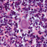
Sporadic Creutzfeldt-Jakob disease: Real-Time Quaking Induced Conversion (RT-QuIC) assay represents a major diagnostic advance

Accepted: 7 September 2021
HTML: 24
All claims expressed in this article are solely those of the authors and do not necessarily represent those of their affiliated organizations, or those of the publisher, the editors and the reviewers. Any product that may be evaluated in this article or claim that may be made by its manufacturer is not guaranteed or endorsed by the publisher.
Authors
Sporadic Creutzfeldt-Jakob disease (sCJD) is a rare and fatal neurodegenerative disorder with an incidence of 1.5 to 2 cases per million population/year. The disease is caused by a proteinaceous infectious agent, named prion (or PrPSc), which arises from the conformational conversion of the cellular prion protein (PrPC). Once formed, PrPSc interacts with the normally folded PrPC coercing it to undergo similar structural rearrangement. The disease is highly heterogeneous from a clinical and neuropathological point of view. The origin of this variability lies in the aberrant structures acquired by PrPSc. At least six different sCJD phenotypes have been described and each of them is thought to be caused by a peculiar PrPSc strain. Definitive sCJD diagnosis requires brain analysis with the aim of identifying intracerebral accumulation of PrPSc which currently represents the only reliable biomarker of the disease. Clinical diagnosis of sCJD is very challenging and is based on the combination of several clinical, instrumental and laboratory tests representing surrogate disease biomarkers. Thanks to the advent of the ultrasensitive Real-Time Quaking-Induced Conversion (RT-QuIC) assay, PrPSc was found in several peripheral tissues of sCJD patients, sometimes even before the clinical onset of the disease. This discovery represents an important step forward for the clinical diagnosis of sCJD. In this manuscript, we present an overview of the current applications and future perspectives of RT-QuIC in the field of sCJD diagnosis.
How to Cite

This work is licensed under a Creative Commons Attribution-NonCommercial 4.0 International License.
PAGEPress has chosen to apply the Creative Commons Attribution NonCommercial 4.0 International License (CC BY-NC 4.0) to all manuscripts to be published.
Similar Articles
- Hao Li, Junliang Chen, Wenjun You, Yizhen Xu, Yaqiong Ye, Haiquan Zhao, Junxing Li, Hui Zhang, Developmental characteristics of cutaneous telocytes in late embryos of the silky fowl , European Journal of Histochemistry: Vol. 68 No. 4 (2024)
- Renata Tabola, Magdalena Zaremba-Czogalla, Dagmara Baczynska, Roberto Cirocchi, Kamila Stach, Krzysztof Grabowski, Katarzyna Augoff, Fibroblast activating protein-α expression in squamous cell carcinoma of the esophagus in primary and irradiated tumors: the use of archival FFPE material for molecular techniques , European Journal of Histochemistry: Vol. 61 No. 2 (2017)
- Chiara Rita Inguscio, Flavia Carton, Barbara Cisterna, Manuela Rizzi, Francesca Boccafoschi, Gabriele Tabaracci, Manuela Malatesta, Low ozone concentrations do not exert cytoprotective effects on tamoxifen-treated breast cancer cells in vitro , European Journal of Histochemistry: Vol. 68 No. 3 (2024)
- Liying Shen, Kongjie Lu, Zhenfeng Chen, Yingwei Zhu, Cong Zhang, Li Zhang, Pre-treatment with galectin-1 attenuates lipopolysaccharide-induced myocarditis by regulating the Nrf2 pathway , European Journal of Histochemistry: Vol. 67 No. 4 (2023)
- A. Mangas, J. Yajeya, N. González, S. Duleu, M. Geffard, R. Coveñas, NO-tryptophan: a new small molecule located in the rat brain , European Journal of Histochemistry: Vol. 60 No. 3 (2016)
- M. Malatesta, M. Giagnacovo, M. Costanzo, B. Conti, I. Genta, R. Dorati, V. Galimberti, M. Biggiogera, C. Zancanaro, Diaminobenzidine photoconversion is a suitable tool for tracking the intracellular location of fluorescently labelled nanoparticles at transmission electron microscopy , European Journal of Histochemistry: Vol. 56 No. 2 (2012)
- E. I. Cortés-Gutiérrez, M. I. Dávila-RodrÃguez, J. L. Fernández, C. López-Fernández, J. Gosálvez, Koilocytes are enriched for alkaline-labile sites , European Journal of Histochemistry: Vol. 54 No. 4 (2010)
- Carlo Pellicciari, Histochemistry in biology and medicine: a message from the citing journals , European Journal of Histochemistry: Vol. 59 No. 4 (2015)
- M. Miko, L. Danisovic, A. Majidi, I. Varga, Ultrastructural analysis of different human mesenchymal stem cells after in vitro expansion: a technical review , European Journal of Histochemistry: Vol. 59 No. 4 (2015)
- E. Guerriero, M. Accardo, F. Capone, G. Colonna, G. Castello, S. Costantini, Assessment of the Selenoprotein M (SELM) over-expression on human hepatocellular carcinoma tissues by immunohistochemistry , European Journal of Histochemistry: Vol. 58 No. 4 (2014)
<< < 27 28 29 30 31 32 33 34 35 36 > >>
You may also start an advanced similarity search for this article.

 https://doi.org/10.4081/ejh.2021.3298


