Morphological and histochemical characterization of the secretory epithelium in the canine lacrimal gland
Submitted: 31 August 2021
Accepted: 14 October 2021
Published: 2 November 2021
Abstract Views: 1141
PDF: 544
HTML: 20
Publisher's note
All claims expressed in this article are solely those of the authors and do not necessarily represent those of their affiliated organizations, or those of the publisher, the editors and the reviewers. Any product that may be evaluated in this article or claim that may be made by its manufacturer is not guaranteed or endorsed by the publisher.

Authors

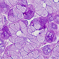
In the present study, the expression of secretory components and vesicular transport proteins in the canine lacrimal gland was examined and morphometric analysis was performed. The secretory epithelium consists of two types of secretory cells with different morphological features. The secretory cells constituting acinar units (type A cells) exhibited higher levels of glycoconjugates, including β-GlcNAc, than the other cell type constituting tubular units (type T cells). Immunoblot analysis revealed that antimicrobial proteins, such as lysozyme, lactoferrin and lactoperoxidase, Rab proteins (Rab3d, Rab27a and Rab27b) and soluble N-ethylmaleimide-sensitive fusion protein attachment protein receptor (SNARE) proteins (VAMP2, VAMP4, VAMP8, syntaxin-1, syntaxin-4 and syntaxin-6), were expressed at various levels. We immunohistochemically demonstrated that the expression patterns of lysozyme, lactoferrin, Raba27a, Rab27b, VAMP4, VAMP8 and syntaxin-6 differed depending on the secretory cell type. Additionally, in type T cells, VAMP4 was confined to a subpopulation of secretory granules, while VAMP8 was detected in almost all of them. The present study displayed the morphological and histochemical characteristics of the secretory epithelium in the canine lacrimal gland. These findings will help elucidate the species-specific properties of this gland.

Dimensions

Altmetric

PlumX Metrics

Downloads

Download data is not yet available.

Citations

Fullard RJ, Snyder C. Protein levels in nonstimulated and stimulated tears of normal human subjects. Invest Ophthalmol Vis Sci 1990;31:1119-26.
Hodges RR, Dartt DA. Regulatory pathways in lacrimal gland epithelium. Int Rev Cytol 2003;231:129-96.
Sack RA, Conradi L, Krumholz D, Beaton A, Sathe S, Morris C. Membrane array characterization of 80 chemokines, cytokines, and growth factors in open- and closed-eye tears: angiogenin and other defense system constituents. Invest Ophthalmol Vis Sci 2005;46:1228-38.
Zhou L, Zhao SZ, Koh SK, Chen L, Vaz C, Tanavde V, et al. In-depth analysis of the human tear proteome. J Proteomics 2012;75:3877–85.
Grosshans BL, Ortiz D, Novick P. Rabs and their effectors: achieving specificity in membrane traffic. Proc Natl Acad Sci USA 2006;103:11821-7.
Williams JA, Chen X, Sabbatini ME. Small G proteins as key regulators of pancreatic digestive enzyme secretion. Am J Physiol Endocrinol Metab 2009;296:E405-15.
Chen X, Edwards JAS, Logsdon CD, Ernst SA, Williams JA. Dominant negative Rab3D inhibits amylase release from mouse pancreatic acini. J Biol Chem 2002;20:18002-9.
Hou Y, Ernst SA, Stuenkel EL, Lentz SI, Williams JA. Rab27A is present in mouse pancreatic acinar cells and is required for digestive enzyme secretion. PloS One 2015;10:e0125596.
Hou Y, Ernst SA, Lentz SI, Williams JA. Genetic deletion of Rab27B in pancreatic acinar cells affects granules size and has inhibitory effects on amylase secretion. Biochem Biophys Res Commun 2016;471:610-5.
Ohnishi H, Ernst SA, Wys N, McNiven M, Williams JA. Rab3D localizes to zymogen granules in rat pancreatic acini and other exocrine glands. Am J Physiol 1996;271:G531-8.
Raffaniello RD, Lin J, Schwimmer R, Ojakian GK. Expression and localization of Rab3D in rat parotid gland. Biochim Biophys Acta 1999;1450:352-63.
Imai A, Yoshie S, Nashida T, Shimomura H, Fukuda M. The small GTPase Rab27B regulates amylase release from rat parotid acinar cells. J Cell Sci 2004;117:1945-53.
Wang Y, Jerdeva G, Yarber FA, da Costa SR, Xie J, Qian L, et al. Cytoplasmic dynein participates in apically targeted stimulated secretory traffic in primary rabbit lacrimal acinar epithelial cells. J Cell Sci 2003;116:2051-65.
Evans E, Zhang W, Jerdeva G, Chen CY, Chen X, Hamm-Alvarez SF, et al. Direct interaction between Rab3D and the polymeric immunoglobulin receptor and trafficking through regulated secretory vesicles in lacrimal gland acinar cells. Am J Cell Physiol 2008;294:C662-74.
Chiang L, Ngo J, Schechter JE, Karvar S, Tolmachova T, Seabra MC, et al. Rab27b regulates exocytosis of secretory vesicles in acinar epithelial cells from the lacrimal gland. Am J Cell Physiol 2011;301:C507-21.
Meng Z, Edman MC, Hsueh PY, Chen CY, Klinngam W, Tolmachova T, et al. Imbalanced Rab3D versus Rab27 increases cathepsin S secretion from lacrimal acini in a mouse model of Sjögren’s Syndrome. Am J Cell Physiol 2016;310:C942-54.
Söllner T, Whiteheart SW, Brunner M, Erdjument-Bromage H, Geromanos S, Tempst P, et al. SNAP receptors implicated in vesicle targeting and fusion. Nature 1993;362:318-24.
Rothman JE. Mechanisms of intracellular protein transport. Nature 1994; 372:55-63.
Rothman JE, Warren G. Implications of the SNARE hypothesis for intracellular membrane topology and dynamics. Curr Biol 1994;4:220-33.
Imai A, Nashida T, Yoshie S, Shimomura H. Intracelular localization of SNARE proteins in rat parotid acinar cells: SNARE complexes on the apical plasma membrane. Arch Oral Biol 2003;48:597-604.
Wang CC, Ng CP, Lu L, Atlashkin V, Zhang W, Seet LF, et al. A role of VAMP8/endobrevin in regulated exocytosis of pancreatic acinar cells. Dev Cell 2004;7:359-71.
Wang CC, Shi H, Guo K, Ng CP, Li J, Gan BQ, et al. VAMP8/endobrevin as a general vesicular SNARE for regulated exocytosis of the exocrine system. Mol Biol Cell 2007;18:1056-63.
Wu K, Jerdeva GV, da Costa SR, Sou E, Schechter JE, Hamm-Alvarez SF. Molecular mechanisms of lacrimal acinar secretory vesicle exocytosis. Exp Eye Res 2006;83:84-96.
Weng N, Thomas DD, Groblewski GE. Pancreatic acinar cells express vesicle-associated membrane protein 2- and 8-specific populations of zymogen granules with distinct and overlapping roles in secretion. J Biol Chem 2007;282:9635-45.
Stoeckelhuber M, Scherer EQ, Janssen KP, Slotta-Huspenina J, Loeffelbein DJ, Rohleder NH, et al. The human submandibular gland: immunohistochemical analysis of SNAREs and cytoskeletal proteins. J Histochem Cytochem 2012;60:110-20.
Gomi H, Osawa H, Uno R, Yasui T, Hosaka M, Torii S, et al. Canine salivary glands: analysis of Rab and SNARE protein expression and SNARE complex formation with diverse tissue properties. J Histochem Cytochem 2017;65:637-53.
Yasui T, Gomi H, Kitahara T, Tsukise A. Ultrastructure and immunohistochemical characterization of proteins concerned with the secretory machinery in goat ceruminous glands. Eur J Histochem 2017;61:222-30.
Dellmann HD. Eye and ear. In: Dellmann HD, Brown EM, Editors. Textbook of veterinary histology. 2nd ed. Philadelphia: Lea and Febiger; 1981. p. 412-40.
Raskin RE. Eyes and adnexa. In: Raskin RE, Meyer DJ, Editors. Atlas of canine and feline cytology. Philadelphia: W.B. Saunders; 2001. p. 367-84.
Martin CL, Munnell J, Kaswan R. Normal ultrastructure and histochemical characteristics of canine lacrimal glands. Am J Vet Res 1988;49:1566-72.
Kuhnel W. [Vergleichende histologische, histochemische und electronenmikroskopische Untersuchungen an Tranedrusen 4 hund].[Article in German]. Z Zellforsch 1968;88:23-38.
Luft JH. Improvements in epoxy resin embedding methods. J Biophys Biochem Cytol 1961;9:409-14.
Newman GR, Jasani B, Williams ED. A simple post-embedding system for the rapid demonstration of tissue antigens under the electron microscope. Histochem J 1983;15:543-55.
Spicer SS, Schulte BA. Diversity of cell glycoconjugates shown histochemically: a perspective. J Histochem Cytochem 1992;40:1-38.
Danguy A. Perspectives in modern glycohistochemistry. Eur J Histochem 1995;39:5-14.
Yamada K. Histochemistry of carbohydrates as performed by physical development procedures. Histochem J 1993;25:95-106.
Gomi H, Mori K, Itohara S, Izumi T. Rab27b is expressed in a wide range of exocytic cells and involved in the delivery of secretory granules near the plasma membrane. Mol Biol Cell 2007;18:4377-86.
Ohashi Y, Dogru M, Tsubota K. Laboratory findings in tear fluid analysis. Clin Chim Acta 2006;369:17-28.
Gipson IK. Distribution of mucins at the ocular surface. Exp Eye Res 2004;78:379-88.
Paulsen F. Cell and molecular biology of human lacrimal gland and nasolacrimal duct mucins. Int Rev Cytol 2006;249:229-79.
Pflugfelder SC, Stern ME. Biological functions of tear film. Exp Eye Res 2020;197:108115.
Gelatt KN. Canine lacrimal gland and nasolacrimal disease. In: Gelatt KN, Editor. Veterinary ophthalmology. Philadelphia: Lea and Febiger; 1991. p. 276-89.
Habata I, Yasui T, Tsukise A. Histochemistry of sialoglycoconjugates in goat submandibular glands. Anat Histol Embryol 2011;40:187-95.
Habata I, Yasui T, Fujimori A, Meyer W, Tsukise A. Histochemical analyses of glycoconjugates and antimicrobial substances in goat labial glands. Acta Histochem 2012;114:454-62.
Sugiura Y, Soeta S, Ichihara T, Nishita M, Murakami M, Amasaki H, et al. Immunohistolocalization and gene expression of the carbonic anhydrase isoenzyme (CA-II and CA-VI) in glands associated with the canine lacrimal apparatus. Anat Histol Embryol 2010;39:1-6.
Haynes RJ, Tighe PJ, Dua HS. Antimicrobial defensin peptides of human ocular surface. Br J Ophthalmol 1999;83:737-41.
Li N, Wang N, Zheng J, Liu XM, Lever OW, Erickson PM, et al. Characterization of human tear proteome using multiple proteomic analysis techniques. J Proteome Res 2005;4:2052-61.
Imai A, Nashida T, Yoshie S, Shimomura H, Fukuda M. Functional involvement of Noc2, a Rab27 effector, in rat parotid acinar cells. Arch Biochem Biophys 2006;455:127-35.
Imai A, Yoshie S, Nashida T, Fukuda M, Shimomura H. Redistribution of small GTP-binding protein, Rab27B, in rat parotid acinar cells after stimulation with isoproterenol. Eur J Oral Sci 2009;117:224-30.
Riedel D, Antonin W, Fernandez-Chacon R, Alvarez de Toledo G, Jo T, Geppert M, et al. Rab3D is required for exocrine exocytosis but for maintenance of normally sized secretory granules. Mol Cell Biol 2002;22:6487-97.
Söllner T, Bennett MK, Whiteheart SW, Scheller RH, Rothman JE. A protein assembly-disassembly pathway in vitro that may correspond to sequential steps of synaptic vesicle docking, activation, and fusion. Cell 1993;75:409-18.
Steegmaier M, Klumperman J, Foletti DL, Yoo JS, Scheller RH. Vesicle-associated membrane protein 4 is implicated in trans-Golgi network vesicle trafficking. Mol Biol Cell 1999;10:1957-72.
Katsumata O, Fujita-Yoshigaki J, Hara-Yokoyama M, Yanagishita M, Furuyama S, Sugiya H. Syntaxin6 separates from GM1a-rich membrane microdomain during granule maturation. Biochem Biophys Res Commun 2007;357:1071-7.
Yokoyama M, Katsumata-Kato O, Fujita-Yoshigaki J. Syntaxin 6 is involved in the maintenance of secretory granules in parotid acinar cells. Int J Oral-Med Sci 2017;15:67-73.
Wong SH, Zhang T, Xu Y, Subramaniam VN, Griffiths G, Hong W. Endobrevin, a novel synaptobrevin/VAMP-like protein preferentially associated with the early endosome. Mol Biol Cell 1998;9:1549-63.
Advani RJ, Bae HR, Bock JB, Chao DS, Prekeris R, Yoo JS, et al. Seven novel mammalian SNARE proteins localize to distinct membrane compartments. J Biol Chem 1998;273:10317-24.
Paumet F, Le Mao J, Martin S, Galli T, David B, Blank U, et al. Soluble NSF attachment protein receptors (SNAREs) in RBL-2H3 mast cells: functional role of syntaxin 4 in exocytosis and identification of a vesicle-associated membrane protein 8-containing secretory compartment. J Immunol 2000;164:5850-7.
Polgar J, Chung SH, Reed GL. Vesicle-associated membrane protein 3 (VAMP-3) and VAMP-8 are present in human platelets and are required for granule secretion. Blood 2002;100:1081-3.
Kamoi M, Ogawa Y, Nakamura S, Dogru M, Nagai T, Obata H, et al. Accumulation of secretory vesicles in the lacrimal gland epithelia is related to non-Sjögren’s type dry eye in visual display terminal users. PLoS One 2012;7:e43688.
Fukui M, Ogawa Y, Mukai S, Kamoi M, Asato T, Kawakami Y, et al. Reduced expression of VAMP8 in lacrimal gland affected by chronic graft-versus-host disease. J Ophthalmol 2017;2017:1639012.
Miyazaki T, Fujiki T, Inoue Y, Takano K. Immunoelectron microscopic identification of lysozyme-expressing cells in human labial salivary glands. Arch Histol Cytol 1998;61:199-214.
Obata H. Anatomy and histopathology of the human lacrimal gland. Cornea 2006;25:S82-9.
Ofri R, Orgad K, Kass PH, Dikstein S. Canine meibometry: establishing baseline values for meibomian gland secretions in dogs. Vet J 2007;174:536-40.

How to Cite

Yasui, T., Miyata, K., Nakatsuka, C., Tsukise, A., & Gomi, H. (2021). Morphological and histochemical characterization of the secretory epithelium in the canine lacrimal gland. European Journal of Histochemistry, 65(4). https://doi.org/10.4081/ejh.2021.3320

Similar Articles

<< < 1 2 3 4 5 6 7 8 9 10 > >> 

You may also start an advanced similarity search for this article.

Publication Facts

Metric
This article
Other articles
Peer reviewers 
2
2.4

Reviewer profiles  N/A

Author statements

Author statements
This article
Other articles
Data availability 
N/A
16%
External funding 
N/A
32%
Competing interests 
N/A
11%
Metric
This journal
Other journals
Articles accepted 
57%
33%
Days to publication 
62
145

Indexed in

Editor & editorial board
profiles
Academic society 
N/A