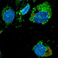
Autofluorescence in freshly isolated adult human liver sinusoidal cells

Lysosomal labelling with mouse anti-human LAMP-1 antibody (green) shows that the autofluorescent material (red) is located within LAMP-1 positive vesicles (arrow heads)
Submitted: 5 October 2021
Accepted: 1 December 2021
Published: 13 December 2021
Abstract Views: 1288
PDF: 664
HTML: 50
Publisher's note
All claims expressed in this article are solely those of the authors and do not necessarily represent those of their affiliated organizations, or those of the publisher, the editors and the reviewers. Any product that may be evaluated in this article or claim that may be made by its manufacturer is not guaranteed or endorsed by the publisher.

Authors

Autofluorescent granules of various sizes were observed in primary human liver endothelial cells (LSECs) upon laser irradiation using a wide range of wavelengths. Autofluorescence was detected in LAMP-1 positive vesicles, suggesting lysosomal location. Confocal imaging of freshly prepared cultures and imaging flow cytometry of non-cultured cells revealed fluorescence in all channels used. Treatment with a lipofuscin autofluorescence quencher reduced autofluorescence, most efficiently in the near UV-area. These results, combined with the knowledge of the very active blood clearance function of LSECs support the notion that lysosomally located autofluorescent material reflected accumulation of lipofuscin in the intact liver. These results illustrate the importance of careful selection of fluorophores, especially when labelling of live cells where the quencher is not compatible.

Dimensions

Altmetric

PlumX Metrics

Downloads

Download data is not yet available.

Citations

Croce AC, De Simone U, Freitas I, Boncompagni E, Neri D, Cillo U, et al. Human liver autofluorescence: an intrinsic tissue parameter discriminating normal and diseased conditions. Lasers Surg Med 2010;42:371-8. DOI: https://doi.org/10.1002/lsm.20923
Shang L, Hosseini M, Liu X, Kisseleva T, Brenner DA. Human hepatic stellate cell isolation and characterization. J Gastroenterol 2018;53:6-17. DOI: https://doi.org/10.1007/s00535-017-1404-4
Brunk UT, Terman A. Lipofuscin: mechanisms of age-related accumulation and influence on cell function. Free Radic Biol Med 2002;33:611-9. DOI: https://doi.org/10.1016/S0891-5849(02)00959-0
Robbins S, Contran RS, Kumar V. Pathologic basis of disease. 5th ed. W.B. Sounders Company; 1994.
Josefsen TD, Sørensen KK, Mørk T, Mathiesen SD, Ryeng KA. Fatal inanition in reindeer (Rangifer tarandus tarandus): pathological findings in completely emaciated carcasses. Acta Vet Scand 2007;49:1-11. DOI: https://doi.org/10.1186/1751-0147-49-27
Croce AC, Bottiroli G. Autofluorescence spectroscopy and imaging: a tool for biomedical research and diagnosis. Eur J Histochem 2014;58;2461. DOI: https://doi.org/10.4081/ejh.2014.2461
Elvevold K, Smedsrød B, Martinez I. The liver sinusoidal endothelial cell: a cell type of controversial and confusing identity. Am J Physiol Gastrointest Liver Physiol 2008;294:G391-G400. DOI: https://doi.org/10.1152/ajpgi.00167.2007
Sørensen KK, McCourt P, Berg T, Crossley C, Couteur DL, Wake K, et al. The scavenger endothelial cell: a new player in homeostasis and immunity. Am J Physiol Regul Integr Comp Physiol 2012;303:R1217-R30. DOI: https://doi.org/10.1152/ajpregu.00686.2011
Sørensen KK, Simón-Santamaría J, McCuskey RS, Smedsrød B. Liver sinusoidal endothelial cells. Compr Physiol 2015;5:1751-74. DOI: https://doi.org/10.1002/cphy.c140078
Croce AC, Ferrigno A, Vairetti M, Bertone R, Freitas I, Bottiroli G. Autofluorescence properties of isolated rat hepatocytes under different metabolic conditions. Photochem Photobiol Sci 2004;3:920-6. DOI: https://doi.org/10.1039/b407358d
Wake K. “Sternzellen” in the liver: perisinusoidal cells with special reference to storage of vitamin A. Am J Anat 1971;132:429-61. DOI: https://doi.org/10.1002/aja.1001320404
LeCluyse EL, Alexandre E. Isolation and culture of primary hepatocytes from resected human liver tissue. In: P. Maurel, editor. Hepatocytes. Totowa: Humana Press; 2010. p. 57-82. DOI: https://doi.org/10.1007/978-1-60761-688-7_3
Kuo J. Electron microscopy: methods and protocols. Berlin: Springer; 2007. DOI: https://doi.org/10.1007/978-1-59745-294-6
Bhandari S, Larsen AK, McCourt P, Smedsrød B, Sørensen, KK. The scavenger funtion of liver sinusoidal endothelial cells in health and disease. Front Physiol 2021;12:e757469. DOI: https://doi.org/10.3389/fphys.2021.757469
Martens JH, Kzhyshkowska J, Falkowski‐Hansen M, Schledzewski K, Gratchev A, Mansmann U, et al. Differential expression of a gene signature for scavenger/lectin receptors by endothelial cells and macrophages in human lymph node sinuses, the primary sites of regional metastasis. J Pathol 2006;208:574-89. DOI: https://doi.org/10.1002/path.1921
Mousavi SA, Sporstøl M, Fladeby C, Kjeken R, Barois N, Berg T. Receptor‐mediated endocytosis of immune complexes in rat liver sinusoidal endothelial cells is mediated by FcγRIIb2. Hepatology 2007;46:871-84. DOI: https://doi.org/10.1002/hep.21748
Strauss O, Phillips A, Ruggiero K, Bartlett A, Dunbar PR. Immunofluorescence identifies distinct subsets of endothelial cells in the human liver. Sci Rep 2017;7:1-13. DOI: https://doi.org/10.1038/srep44356
D'Ambrosio DN, Walewski JL, Clugston RD, Berk PD, Rippe RA, Blaner WS. Distinct populations of hepatic stellate cells in the mouse liver have different capacities for retinoid and lipid storage. PloS One 2011;6:e24993. DOI: https://doi.org/10.1371/journal.pone.0024993
Le Couteur DG, Warren A, Cogger VC, Smedsrød B, Sørensen KK, De Cabo R, et al. Old age and the hepatic sinusoid. Anat Rec 2008;291:672-83. DOI: https://doi.org/10.1002/ar.20661
Werner M, Driftmann S, Kleinehr K, Kaiser GM, Mathé Z, Treckmann J-W, et al. All-in-one: advanced preparation of human parenchymal and non-parenchymal liver cells. PloS One 2015;10:e0138655. DOI: https://doi.org/10.1371/journal.pone.0138655
Armstrong D, Koppang N. Ceroid-lipofuscinosis: a model for aging. In: RS Sohal, editor. Age pigments. Amsterdam: Elsevier; 1981. p. 355-82.
Reddy JK, Lalwani ND, Reddy MK, Qureshi SA. Excessive accumulation of autofluorescent lipofuscin in the liver during hepatocarcinogenesis by methyl clofenapate and other hypolipidemic peroxisome proliferators. Canc Res 1982;42:259-66.
Ettinger A, Wittmann T. Fluorescence live cell imaging. Methods Cell Biol 2014;123:77-94. DOI: https://doi.org/10.1016/B978-0-12-420138-5.00005-7

Supporting Agencies

Norwegian Research Council
Kjetil Elvevold, Department of Medical Biology, UiT – The Arctic University of Norway, Tromsø

Current address:
The Norwegian Institute of Food, Fisheries and Aquaculture Research, Tromsø, Norway

How to Cite

Larsen, A. K., Simón-Santamaría, J., Elvevold, K., Ericzon, B. G. ., Mortensen, K. E., McCourt, P., … Sørensen, K. K. (2021). Autofluorescence in freshly isolated adult human liver sinusoidal cells. European Journal of Histochemistry, 65(4). https://doi.org/10.4081/ejh.2021.3337

Similar Articles

1 2 3 4 5 6 7 8 9 10 > >> 

You may also start an advanced similarity search for this article.