Ghrelin reduces cerebral ischemic injury in rats by reducing M1 microglia/macrophages

Submitted: 21 October 2021
Accepted: 18 December 2021
Published: 12 January 2022
Abstract Views: 1281
PDF: 562
HTML: 21
Publisher's note
All claims expressed in this article are solely those of the authors and do not necessarily represent those of their affiliated organizations, or those of the publisher, the editors and the reviewers. Any product that may be evaluated in this article or claim that may be made by its manufacturer is not guaranteed or endorsed by the publisher.

Authors

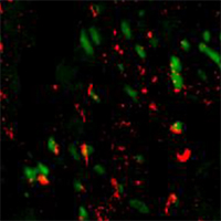
The purpose of this study was to investigate the effect of Ghrelin on the polarization of microglia/ macrophages after cerebral ischemia (CI) in rats. 60 wild-type SD rats were randomly divided into sham group, CI group, CI+Ghrelin group, 20 rats in each group. The modified Longa suture method was used to establish the middle cerebral artery occlusion (MCAO) model in rats. Before surgery, Ghrelin was injected subcutaneously (100μg/kg, twice a day) for 4 consecutive weeks. After modeling, neurological function scores were performed with three behavioral experiments: mNSS score, Corner test, and Rotarod test, to evaluate the recovery of neurological function after Ghrelin treatment. At the same time, the brain tissues were collected and stained with 2,3,5-triphenyltetrazolium chloride (TTC) to detect the cerebral infarct volume. RT-qPCR was used to detect the expression of TNF-α and IL-1β in the ischemic brain tissue, and the TUNEL staining was used to detect the apoptosis of brain tissue. Flow cytometry was used to detect the percentage of M1 type microglia/macrophages which were isolated by trypsin digestion of fresh cerebral cortex. Then, the Western blotting and immunofluorescence method were used to detect the phosphorylation level of AKT (P-AKT) and AKT. Compared with the CI group, the neurological function of the rats in the CI+Ghrelin group was dramatically improved, and the cerebral infarction area was dramatically reduced. At the same time, the expression of TNF-α and IL-1β in the ischemic brain tissue of rats in the CI+Ghrelin group decreased, and the apoptotic cells in the brain tissue also decreased. Compared with the CI treatment group, the activation of M1 microglia/macrophages in the cortex of the ischemic side of the infarct and the peri-infarct area in the CI+Ghrelin group was dramatically inhibited. At the same time, the ratio of P-AKT/AKT of the brain tissue in the CI+Ghrelin group was dramatically higher than that of the CI group. In the rat cerebral ischemia model, Ghrelin can promote the repair of brain damage and the recovery of neurological function after ischemia. Its mechanism may be related to activating AKT to selectively reduce M1 microglia/macrophages, reducing inflammation and cell apoptosis in brain tissue.

Dimensions

Altmetric

PlumX Metrics

Downloads

Download data is not yet available.

Citations

Wu S, Wu B, Liu M, Chen Z, Wang W, Anderson CS et al. Stroke in China: advances and challenges in epidemiology, prevention, and management. Lancet Neurol 2019;18:394-405. DOI: https://doi.org/10.1016/S1474-4422(18)30500-3
Wardlaw JM, Murray V, Berge E, Del ZG. Thrombolysis for acute ischaemic stroke. Cochrane Database Syst Rev 2014;(7):D213. DOI: https://doi.org/10.1002/14651858.CD000213.pub3
Garber C, Soung A, Vollmer LL, Kanmogne M, Last A, Brown J, et al. T cells promote microglia-mediated synaptic elimination and cognitive dysfunction during recovery from neuropathogenic flaviviruses. Nat Neurosci 2019;22:1276-88. DOI: https://doi.org/10.1038/s41593-019-0427-y
Cengiz P, Zafer D, Chandrashekhar JH, Chanana V, Bogost J, Waldman A, et al. Developmental differences in microglia morphology and gene expression during normal brain development and in response to hypoxia-ischemia. Neurochem Int 2019;127:137-47. DOI: https://doi.org/10.1016/j.neuint.2018.12.016
Tsuda M. Microglia-mediated regulation of neuropathic pain: Molecular and cellular mechanisms. Biol Pharm Bull 2019;42:1959-68. DOI: https://doi.org/10.1248/bpb.b19-00715
Przanowski P, Mondal SS, Cabaj A, Debski KJ, Wojtas B, Gielniewski B, et al. Open chromatin landscape of rat microglia upon proinvasive or inflammatory polarization. Glia 2019;67:2312-28. DOI: https://doi.org/10.1002/glia.23686
Cherry JD, Olschowka JA, O'Banion MK. Neuroinflammation and M2 microglia: the good, the bad, and the inflamed. J Neuroinflammation 2014;11:98. DOI: https://doi.org/10.1186/1742-2094-11-98
Tang Z, Gan Y, Liu Q, Yin JX, Liu Q, Shi J, et al. CX3CR1 deficiency suppresses activation and neurotoxicity of microglia/macrophage in experimental ischemic stroke. J Neuroinflammation 2014;11:26. DOI: https://doi.org/10.1186/1742-2094-11-26
Kang S, Moon NR, Kim DS, Kim SH, Park S. Central acylated ghrelin improves memory function and hippocampal AMPK activation and partly reverses the impairment of energy and glucose metabolism in rats infused with beta-amyloid. Peptides 2015;71:84-93. DOI: https://doi.org/10.1016/j.peptides.2015.07.005
Zeng M, Huang C, Zheng H, Chen Q, He W, Deng Y. Effects of ghrelin on iNOS-derived NO promoted LPS-induced pulmonary alveolar epithelial A549 cells apoptosis. Cell Physiol Biochem 2018;49:1840-55. DOI: https://doi.org/10.1159/000493630
Diano S, Farr SA, Benoit SC, McNay EC, Da SI, Horvath B, et al. Ghrelin controls hippocampal spine synapse density and memory performance. Nat Neurosci 2006;9:381-8. DOI: https://doi.org/10.1038/nn1656
Jeon SG, Hong SB, Nam Y, Tae J, Yoo A, Song EJ, et al. Ghrelin in Alzheimer's disease: Pathologic roles and therapeutic implications. Ageing Res Rev 2019;55:100945. DOI: https://doi.org/10.1016/j.arr.2019.100945
Theodoropoulou A, Metallinos IC, Psyrogiannis A, Vagenakis GA, Kyriazopoulou V. Ghrelin and leptin secretion in patients with moderate Alzheimer's disease. J Nutr Health Aging 2012;16:472-7. DOI: https://doi.org/10.1007/s12603-012-0058-4
Zhou F, Xu J, Ying GY, Wang L, Zhu XD. Ghrelin attenuated cerebral ischemia reperfusion injury in rats. CNS Neurosci Ther 2012;18:945-946. DOI: https://doi.org/10.1111/cns.12006
Chen J, Wang Z, Zheng Z, Chen Y, Khor S, Shi K, et al. Neuron and microglia/macrophage-derived FGF10 activate neuronal FGFR2/PI3K/Akt signaling and inhibit microglia/macrophages TLR4/NF-kappaB-dependent neuroinflammation to improve functional recovery after spinal cord injury. Cell Death Dis 2017;8:e3090. DOI: https://doi.org/10.1038/cddis.2017.490
Hu X, Li P, Guo Y, Wang H, Leak RK, Chen S, et al. Microglia/macrophage polarization dynamics reveal novel mechanism of injury expansion after focal cerebral ischemia. Stroke 2012;43:3063-70. DOI: https://doi.org/10.1161/STROKEAHA.112.659656
Benakis C, Garcia-Bonilla L, Iadecola C, Anrather J. The role of microglia and myeloid immune cells in acute cerebral ischemia. Front Cell Neurosci 2014;8:461. DOI: https://doi.org/10.3389/fnins.2014.00044
Zarruk JG, Greenhalgh AD, David S. Microglia and macrophages differ in their inflammatory profile after permanent brain ischemia. Exp Neurol 2018;301:120-32. DOI: https://doi.org/10.1016/j.expneurol.2017.08.011
Warrington JP, Drummond HA, Granger JP, Ryan MJ. Placental ischemia-induced increases in brain water content and cerebrovascular permeability: role of TNF-alpha. Am J Physiol Regul Integr Comp Physiol 2015;309:R1425-31. DOI: https://doi.org/10.1152/ajpregu.00372.2015
Esenwa CC, Elkind MS. Inflammatory risk factors, biomarkers and associated therapy in ischaemic stroke. Nat Rev Neurol 2016;12:594-604. DOI: https://doi.org/10.1038/nrneurol.2016.125
Zhao SC, Ma LS, Chu ZH, Xu H, Wu WQ, Liu F. Regulation of microglial activation in stroke. Acta Pharmacol Sin 2017;38:445-58. DOI: https://doi.org/10.1038/aps.2016.162

Ethics Approval

This study was approved by the Animal Ethics Committee of the Animal Center of Third Medical Centre Chinese PLA General Hospital (16-PL-EA-3421)

How to Cite

Tian, R. ., & Mao, G. (2022). Ghrelin reduces cerebral ischemic injury in rats by reducing M1 microglia/macrophages. European Journal of Histochemistry, 66(1). https://doi.org/10.4081/ejh.2022.3350