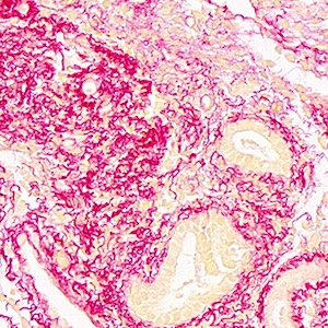
The matrix stiffness is increased in the eutopic endometrium of adenomyosis patients: a study based on atomic force microscopy and histochemistry

Submitted: 15 August 2024
Accepted: 18 November 2024
Published: 4 December 2024
Abstract Views: 473
PDF: 88
HTML: 0
Publisher's note
All claims expressed in this article are solely those of the authors and do not necessarily represent those of their affiliated organizations, or those of the publisher, the editors and the reviewers. Any product that may be evaluated in this article or claim that may be made by its manufacturer is not guaranteed or endorsed by the publisher.

Authors

Previous ultrasound studies suggest that patients with adenomyosis (AM) exhibit increased uterine cavity stiffness, although direct evidence regarding extracellular matrix (ECM) content and its specific impact on endometrial stiffness remains limited. This study utilized atomic force microscopy to directly measure endometrial stiffness and collagen morphology, enabling a detailed analysis of the endometrium’s mechanical properties: through this approach, we established direct evidence of increased endometrial stiffness and fibrosis in patients with AM. Endometrial specimens were also stained with Picrosirius red or Masson’s trichrome to quantify fibrosis, and additional analyses assessed α-SMA and Ki-67 expression. Studies indicate that pathological conditions significantly influence the mechanical properties of endometrial tissue. Specifically, adenomyotic endometrial tissue demonstrates increased stiffness, associated with elevated ECM and fibrosis content, whereas normal endometrial samples are softer with lower ECM content. AM appears to alter both the mechanical and histological characteristics of the eutopic endometrium. Higher ECM content may significantly impact endometrial mechanical properties, potentially contributing to AM-associated decidualization defects and fertility challenges.

Dimensions

Altmetric

PlumX Metrics

Downloads

Download data is not yet available.

Citations

1. Vannuccini S, Tosti C, Carmona F, Huang SJ, Chapron C, Guo S-W, et al. Pathogenesis of adenomyosis: an update on molecular mechanisms. Reprod Biomed Online 2017;35:592-601. DOI: https://doi.org/10.1016/j.rbmo.2017.06.016
2. Vannuccini S, Petraglia F. Recent advances in understanding and managing adenomyosis. F1000Res 2019;8:283-92. DOI: https://doi.org/10.12688/f1000research.17242.1
3. Bergeron C, Amant F, Ferenczy A. Pathology and physiopathology of adenomyosis. Best Pract Res Clin Obstet Gynaecol 2006;20:511-21. DOI: https://doi.org/10.1016/j.bpobgyn.2006.01.016
4. Benagiano G, Habiba M, Brosens I. The pathophysiology of uterine adenomyosis: an update. Fertil Steril 2012;98:572-9. DOI: https://doi.org/10.1016/j.fertnstert.2012.06.044
5. Hufnagel D, Li F, Cosar E, Krikun G, Taylor HS. The role of stem cells in the etiology and pathophysiology of endometriosis. Semin Reprod Med 2015;33:333-40. DOI: https://doi.org/10.1055/s-0035-1564609
6. Liu X, Shen M, Qi Q, Zhang H, Guo S-W. Corroborating evidence for platelet-induced epithelial-mesenchymal transition and fibroblast-to-myofibroblast transdifferentiation in the development of adenomyosis. Hum Reprod 2016;31:734-49. DOI: https://doi.org/10.1093/humrep/dew018
7. Shen M, Liu X, Zhang H, Guo S-W. Transforming growth factor β1 signaling coincides with epithelial-mesenchymal transition and fibroblast-to-myofibroblast transdifferentiation in the development of adenomyosis in mice. Hum Reprod 2016;31:355-69. DOI: https://doi.org/10.1093/humrep/dev314
8. Liu X, Ding D, Ren Y, Guo S-W. Transvaginal elastosonography as an imaging technique for diagnosing adenomyosis. Reprod Sci 2018;25:498-514. DOI: https://doi.org/10.1177/1933719117750752
9. Gadegaard N. Atomic force microscopy in biology: technology and techniques. Biotech Histochem 2006;81:87-97. DOI: https://doi.org/10.1080/10520290600783143
10. Allison DP, Mortensen NP, Sullivan CJ, Doktycz MJ. Atomic force microscopy of biological samples. Wiley Interdiscip Rev Nanomed Nanobiotechnol 2010;2:618-34. DOI: https://doi.org/10.1002/wnan.104
11. Stylianou A, Kontomaris S-V, Grant C, Alexandratou E. Atomic force microscopy on biological materials related to pathological conditions. Scanning 2019;2019:8452851. DOI: https://doi.org/10.1155/2019/8452851
12. Darling EM, Topel M, Zauscher S, Vail TP, Guilak F. Viscoelastic properties of human mesenchymally-derived stem cells and primary osteoblasts, chondrocytes, and adipocytes. J Biomech 2008;41:454-64. DOI: https://doi.org/10.1016/j.jbiomech.2007.06.019
13. Jiang X, Asbach P, Streitberger K-J, Thomas A, Hamm B, Braun J, et al. In vivo high-resolution magnetic resonance elastography of the uterine corpus and cervix. Eur Radiol 2014;24:3025-33. DOI: https://doi.org/10.1007/s00330-014-3305-8
14. Li QS, Lee GYH, Ong CN, Lim CT. AFM indentation study of breast cancer cells. Biochem Biophys Res Commun 2008;374:609-13. DOI: https://doi.org/10.1016/j.bbrc.2008.07.078
15. Canetta E, Riches A, Borger E, Herrington S, Dholakia K, Adya AK. Discrimination of bladder cancer cells from normal urothelial cells with high specificity and sensitivity: combined application of atomic force microscopy and modulated Raman spectroscopy. Acta Biomater 2014;10:2043-55. DOI: https://doi.org/10.1016/j.actbio.2013.12.057
16. Prabhune M, Belge G, Dotzauer A, Bullerdiek J, Radmacher M. Comparison of mechanical properties of normal and malignant thyroid cells. Micron 2012;43:1267-72. DOI: https://doi.org/10.1016/j.micron.2012.03.023
17. Stolz M, Raiteri R, Daniels AU, VanLandingham MR, Baschong W, Aebi U. Dynamic elastic modulus of porcine articular cartilage determined at two different levels of tissue organization by indentation-type atomic force microscopy. Biophys J 2004;86:3269-83. DOI: https://doi.org/10.1016/S0006-3495(04)74375-1
18. Liang T, Che Y-J, Chen X, Li H-T, Yang H-L, Luo Z-P. Nano and micro biomechanical alterations of annulus fibrosus after in situ immobilization revealed by atomic force microscopy. J Orthop Res 2019;37:232-8. DOI: https://doi.org/10.1002/jor.24168
19. Zuo F, Kaminski N, Eugui E, Allard J, Yakhini Z, Ben-Dor A, et al. Gene expression analysis reveals matrilysin as a key regulator of pulmonary fibrosis in mice and humans. Proc Natl Acad Sci USA 2002;99:6292-7. DOI: https://doi.org/10.1073/pnas.092134099
20. Alonso-Magdalena P, Brössner C, Reiner A, Cheng G, Sugiyama N, Warner M, et al. A role for epithelial-mesenchymal transition in the etiology of benign prostatic hyperplasia. Proc Natl Acad Sci USA 2009;106:2859-63. DOI: https://doi.org/10.1073/pnas.0812666106
21. Pathmanathan N, Balleine RL. Ki67 and proliferation in breast cancer. J Clin Pathol 2013;66:512-6. DOI: https://doi.org/10.1136/jclinpath-2012-201085
22. Bao G, Suresh S. Cell and molecular mechanics of biological materials. Nat Mater 2003;2:715-25. DOI: https://doi.org/10.1038/nmat1001
23. Darling EM, Di Carlo D. High-throughput assessment of cellular mechanical properties. Annu Rev Biomed Eng 2015;17:35-62. DOI: https://doi.org/10.1146/annurev-bioeng-071114-040545
24. Pettenuzzo S, Arduino A, Belluzzi E, Pozzuoli A, Fontanella CG, Ruggieri P, et al. Biomechanics of chondrocytes and chondrons in healthy conditions and osteoarthritis: a review of the mechanical characterisations at the microscale. Biomedicines 2023;11:1942. DOI: https://doi.org/10.3390/biomedicines11071942
25. Charras GT, Horton MA. Single cell mechanotransduction and its modulation analyzed by atomic force microscope indentation. Biophys J 2002;82:2970-81. DOI: https://doi.org/10.1016/S0006-3495(02)75638-5
26. Trache A, Meininger GA. Atomic force microscopy (AFM). Curr Protoc Microbiol 2008;Chapter 2:Unit 2C.2. DOI: https://doi.org/10.1002/9780471729259.mc02c02s8
27. Liu S, Han Y, Kong L, Wang G, Ye Z. Atomic force microscopy in disease-related studies: Exploring tissue and cell mechanics. Microsc Res Tech 2024;87:660-84. DOI: https://doi.org/10.1002/jemt.24471
28. Guolla L, Bertrand M, Haase K, Pelling AE. Force transduction and strain dynamics in actin stress fibres in response to nanonewton forces. J Cell Sci 2012;125:603-13. DOI: https://doi.org/10.1242/jcs.088302
29. Haase K, Pelling AE. Investigating cell mechanics with atomic force microscopy. J R Soc Interface 2015;12:20140970. DOI: https://doi.org/10.1098/rsif.2014.0970
30. Kurek A, Kłosowicz E, Sofińska K, Jach R, Barbasz J. Methods for studying endometrial pathology and the potential of atomic force microscopy in the research of endometrium. Cells 2021;10:219-36. DOI: https://doi.org/10.3390/cells10020219
31. Carvalho L, Podgaec S, Bellodi-Privato M, Falcone T, Abrão MS. Role of eutopic endometrium in pelvic endometriosis. J Minim Invasive Gynecol 2011;18:419-27. DOI: https://doi.org/10.1016/j.jmig.2011.03.009
32. Brosens I, Brosens JJ, Benagiano G. The eutopic endometrium in endometriosis: are the changes of clinical significance? Reprod Biomed Online 2012;24:496-502. DOI: https://doi.org/10.1016/j.rbmo.2012.01.022
33. Ota H, Igarashi S, Hatazawa J, Tanaka T. Is adenomyosis an immune disease? Hum Reprod Update 1998;4:360-7. DOI: https://doi.org/10.1093/humupd/4.4.360
34. Ruiz LA, Báez-Vega PM, Ruiz A, Peterse DP, Monteiro JB, Bracero N, et al. Dysregulation of lysyl oxidase expression in lesions and endometrium of women with endometriosis. Reprod Sci. 2015;22:1496-508. DOI: https://doi.org/10.1177/1933719115585144
35. Grewal S, Carver JG, Ridley AJ, Mardon HJ. Implantation of the human embryo requires Rac1-dependent endometrial stromal cell migration. Proc Natl Acad Sci USA 2008;105:16189-94. DOI: https://doi.org/10.1073/pnas.0806219105
36. Cao J, Li H, Tang H, Gu X, Wang Y, Guan D, et al. Stiff extracellular matrix promotes invasive behaviors of trophoblast cells. Bioengineering (Basel) 2023;10:384-97. DOI: https://doi.org/10.3390/bioengineering10030384
37. Kolácná L, Bakesová J, Varga F, Kostáková E, Plánka L, Necas A, et al. Biochemical and biophysical aspects of collagen nanostructure in the extracellular matrix. Physiol Res 2007;56:S51-S60. DOI: https://doi.org/10.33549/physiolres.931302
38. Xiao Q, Ge G. Lysyl oxidase, extracellular matrix remodeling and cancer metastasis. Cancer Microenviron 2012;5:261-73. DOI: https://doi.org/10.1007/s12307-012-0105-z
39. Bailey AJ. Molecular mechanisms of ageing in connective tissues. Mech Ageing Dev 2001;122:735-55. DOI: https://doi.org/10.1016/S0047-6374(01)00225-1
40. Miles CA, Avery NC, Rodin VV, Bailey AJ. The increase in denaturation temperature following cross-linking of collagen is caused by dehydration of the fibres. J Mol Biol 2005;346:551-6. DOI: https://doi.org/10.1016/j.jmb.2004.12.001
41. Zakaria MA, Aziz J, Rajab NF, Chua EW, Masre SF. Tissue rigidity increased during carcinogenesis of NTCU-induced lung squamous cell carcinoma in vivo. Biomedicines 2022;10:238-50. DOI: https://doi.org/10.3390/biomedicines10102382
42. Achilli M, Mantovani D. Tailoring mechanical properties of collagen-based scaffolds for vascular tissue engineering: the effects of pH, temperature and ionic strength on gelation. Polymers 2010;2:664-80. DOI: https://doi.org/10.3390/polym2040664
43. Arora PD, McCulloch CA. Dependence of collagen remodelling on alpha-smooth muscle actin expression by fibroblasts. J Cell Physiol 1994;159:161-75. DOI: https://doi.org/10.1002/jcp.1041590120
44. Wang J, Fan J, Laschinger C, Arora PD, Kapus A, Seth A, et al. Smooth muscle actin determines mechanical force-induced p38 activation. J Biol Chem 2005;280:7273-84. DOI: https://doi.org/10.1074/jbc.M410819200
45. Altayyeb A, Othman E, Khashbah M, Esmaeel A, El-Mokhtar M, Lambalk C, et al. Characterization of mechanical signature of eutopic endometrial stromal cells of endometriosis patients. Reprod Sci 2020;27:364-74. DOI: https://doi.org/10.1007/s43032-019-00042-3

Ethics Approval

the experimental protocols were approved by the Medical Ethics Review Board of the First Affiliated Hospital of Soochow University, Suzhou, Jiangsu Province, China

Supporting Agencies

National Natural Science Foundation of China

How to Cite

Wang, X., Cai, W., Liang, T., Li, H., Gu, Y., Wei, X., … Yang, X. (2024). The matrix stiffness is increased in the eutopic endometrium of adenomyosis patients: a study based on atomic force microscopy and histochemistry. European Journal of Histochemistry, 68(4). https://doi.org/10.4081/ejh.2024.4131

Similar Articles

1 2 3 4 5 6 7 8 9 10 > >> 

You may also start an advanced similarity search for this article.

Publication Facts

Metric
This article
Other articles
Peer reviewers 
2
2.4

Reviewer profiles  N/A

Author statements

Author statements
This article
Other articles
Data availability 
N/A
16%
External funding 
N/A
32%
Competing interests 
N/A
11%
Metric
This journal
Other journals
Articles accepted 
57%
33%
Days to publication 
110
145

Indexed in

Editor & editorial board
profiles
Academic society 
N/A